66. Cholecystitis
High yield
Gallbladder inflammation, usually from a stuck stone; graded by Tokyo guidelines and treated by early laparoscopic cholecystectomy.
Exam Prep · State examSurgery · HPBSurgery · Emergency and trauma
1. Definition
- Acute cholecystitis → acute inflammation of the gallbladder, usually from obstruction of the cystic duct by a stone
- Acute calculous cholecystitis → 90–95%
- Acalculous cholecystitis → inflammation without stones; critically ill patients (5–10%)
- Chronic cholecystitis → long-standing inflammation and fibrosis from repeated attacks
- Variants → empyema (pus), gangrenous, emphysematous (gas-forming), perforated, xanthogranulomatous
2. Epidemiology
- Acute cholecystitis → develops in about 10–20% of patients with symptomatic gallstones
- Women more than men (calculous); older men in gangrenous and emphysematous forms
- Acalculous → ICU patients, burns, trauma, major surgery, sepsis, TPN; mortality up to 30–50%
- Emphysematous → men, diabetes (about half), vascular disease
- 20% of emergency general surgery admissions in some series relate to gallstones
3. Aetiology and risk factors
- Calculous:
- Stone stuck in Hartmann's pouch or cystic duct
- Risk factors as for gallstones (female, obesity, pregnancy, rapid weight loss)
- Acalculous:
- Critical illness, sepsis, shock, mechanical ventilation
- Burns, major trauma, major surgery (cardiac)
- Total parenteral nutrition, long fasting (stasis)
- Opioids, blood transfusion
- Infections → CMV, Salmonella typhi, HIV-related (cryptosporidium)
- Vasculitis, diabetes
- Emphysematous → Clostridium perfringens, E. coli; ischaemia of the wall
4. Pathogenesis
Calculous
- Stone blocks the cystic duct
- Gallbladder distends; mucosa releases phospholipase A, which turns lecithin into toxic lysolecithin → chemical inflammation
- Wall oedema, raised pressure → venous then arterial compromise
- Secondary bacterial infection (about 50%: E. coli, Klebsiella, Enterococcus, anaerobes)
- Untreated → empyema, gangrene, perforation
- Perforation types (Niemeier):
- I → free perforation, biliary peritonitis
- II → localised, pericholecystic abscess (most common)
- III → cholecystoenteric fistula
- Inflamed gallbladder stuck to the duodenum, colon, omentum → mass
Acalculous
- Stasis (fasting, opioids) and ischaemia (low flow, shock) → concentrated bile injures the wall → early gangrene and perforation
Chronic
- Repeated attacks → thick fibrotic wall, small contracted gallbladder, Rokitansky-Aschoff sinuses
- Calcified wall → porcelain gallbladder
5. Classification
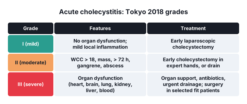
Tokyo Guidelines 2018 (TG18): severity
- Grade I (mild) → no organ dysfunction; mild local inflammation; healthy patient
- Grade II (moderate) → any of:
- White cell count over 18 × 10⁹/L
- Palpable tender RUQ mass
- Symptoms over 72 hours
- Marked local inflammation (gangrene, abscess, emphysematous, biliary peritonitis)
- Grade III (severe) → organ dysfunction: cardiovascular (needs vasopressors), neurological (confusion), respiratory (PaO₂/FiO₂ under 300), renal (oliguria, creatinine over 176 µmol/L), hepatic (INR over 1.5), haematological (platelets under 100 × 10⁹/L)
Tokyo diagnostic criteria
- A Local → Murphy's sign; RUQ mass, pain or tenderness
- B Systemic → fever, raised CRP, raised white cell count
- C Imaging → typical findings
- Suspected → one A + one B; definite → A + B + C
By type
- Calculous, acalculous; acute, chronic; empyema, gangrenous, emphysematous, perforated, xanthogranulomatous
6. Clinical picture
Symptoms
- Constant RUQ pain over 6 hours (unlike colic), worse on breathing and movement
- Radiates to the right shoulder or scapula (diaphragm irritation)
- Nausea, vomiting, anorexia
- Fever, malaise
- Previous attacks of biliary colic
- Elderly and diabetic patients → few symptoms, more gangrene
- Acalculous → unexplained fever, sepsis, rising LFTs in an ICU patient
- Chronic → recurrent episodes of colic, fatty food intolerance, dyspepsia
Signs
- Fever, tachycardia
- RUQ tenderness, guarding
- Murphy's sign → patient stops breathing in when the examiner's fingers under the right costal margin meet the inflamed gallbladder; not seen on the left side (control)
- Boas' sign → hyperaesthesia below the right scapula
- Palpable tender mass (gallbladder with omentum wrapped around it) in about 30%
- Mild jaundice (10%) → oedema near the CBD, Mirizzi, or CBD stone
- Peritonitis → perforation
7. Diagnosis
- Bloods:
- FBC → leucocytosis
- CRP
- LFT → mild rise in bilirubin, ALP; marked rise suggests CBD stone or Mirizzi
- Amylase or lipase → exclude pancreatitis
- U&E, lactate, blood cultures if septic; organ function for Tokyo grading
- Ultrasound → first test:
- Gallstones, stone impacted in the neck
- Thick wall over 4 mm, wall oedema ("double-wall sign")
- Pericholecystic fluid
- Distended gallbladder
- Sonographic Murphy's sign
- HIDA scan (hepatobiliary scintigraphy) → most sensitive and specific: gallbladder does not fill (cystic duct blocked); used if ultrasound unclear
- CT → complications (gangrene, abscess, perforation, gas in the wall in emphysematous), alternative diagnoses
- MRCP → suspected CBD stones
- Chest X-ray, ECG → exclude chest and heart causes; air under the diaphragm
- Plain X-ray → gas in the gallbladder wall (emphysematous), calcified wall (porcelain)
8. Differential diagnosis
- Biliary colic (shorter, no fever, no inflammation)
- Acute cholangitis (jaundice, high fever, rigors)
- Acute pancreatitis
- Perforated peptic ulcer
- Acute appendicitis (high, retrocaecal, in pregnancy)
- Right lower lobe pneumonia, myocardial infarction
- Hepatitis, liver abscess, Fitz-Hugh-Curtis syndrome (gonococcal perihepatitis)
- Right pyelonephritis, renal colic
- Gallbladder cancer
9. Treatment
Conservative
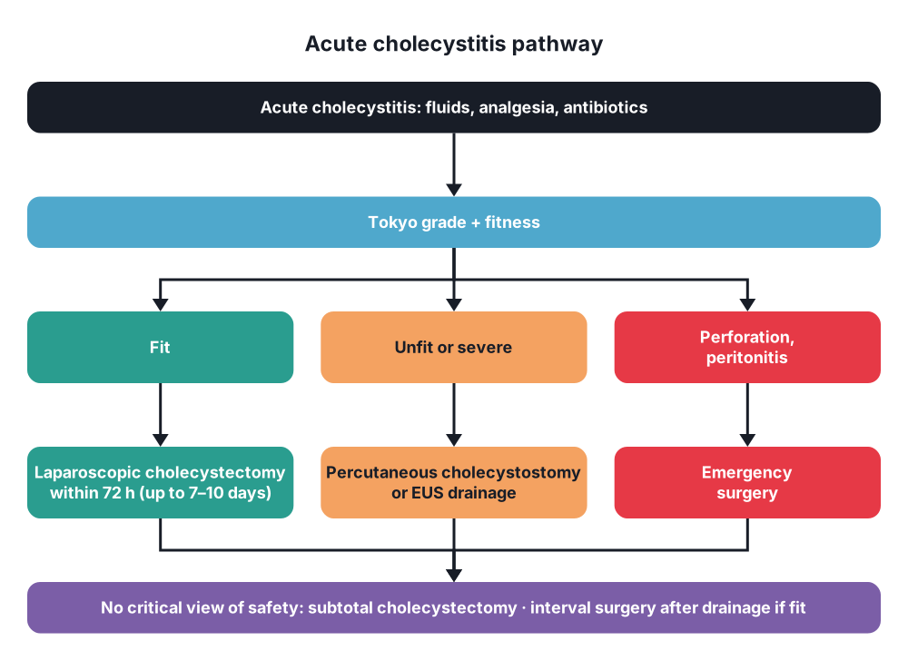
- Initial care (all):
- Admit, nil by mouth or light diet, IV fluids
- Analgesia → NSAIDs, opioids
- Antibiotics → e.g. co-amoxiclav, or cefuroxime with metronidazole, piperacillin-tazobactam for severe (local guidance)
- Antiemetics, VTE prophylaxis
- Assess fitness (Charlson comorbidity index, ASA) and Tokyo grade
- Antibiotics → stop within 24 hours after cholecystectomy for grade I–II (no benefit after)
- Percutaneous cholecystostomy (ultrasound- or CT-guided drain) → unfit patients (grade II not fit, grade III not fit), acalculous cholecystitis, failure of conservative care
- Drain stays 4–6 weeks; tubogram before removal; interval cholecystectomy if fit
- Endoscopic gallbladder drainage → EUS-guided gallbladder stent (lumen-apposing metal stent) or transpapillary stent → for patients unfit for surgery
Surgical
- Early laparoscopic cholecystectomy → standard of care:
- Best within 72 hours of symptoms, but can be done up to 7–10 days (index admission)
- Compared with delayed surgery (6–12 weeks) → shorter total stay, fewer readmissions, same or lower complication rate (CHOCOLATE, ACDC trials; Cochrane)
- Up to 20–30% wait for delayed surgery then return with another attack
- Tokyo-guided plan:
- Grade I → early laparoscopic cholecystectomy
- Grade II → early laparoscopic cholecystectomy in an experienced centre; if not possible or unfit → antibiotics, drainage, then delayed surgery
- Grade III → organ support, antibiotics; early cholecystectomy only in selected fit patients in expert centres; otherwise urgent drainage (cholecystostomy) and later surgery
- Technique notes:
- Decompress a tense gallbladder with a needle first
- Aim for the critical view of safety; if not possible → bail-out (subtotal cholecystectomy, fundus-first, conversion)
- Drain the bed if needed
- Emergency surgery → perforation with peritonitis, emphysematous or gangrenous cholecystitis (with sepsis)
- Acalculous → cholecystostomy first; cholecystectomy if gangrene, perforation or drainage fails
- Chronic cholecystitis → elective laparoscopic cholecystectomy
- Porcelain gallbladder → cholecystectomy (cancer risk, lower than once thought)
10. Complications
- Empyema → gallbladder full of pus; spiking fever, sepsis
- Gangrene and perforation → abscess, biliary peritonitis (mortality up to 30%)
- Cholecystoenteric fistula → gallstone ileus
- Mirizzi syndrome
- Emphysematous → gangrene, perforation; mortality about 15%
- Sepsis, multiorgan failure
- Surgery → bile duct injury (higher in acute inflammation), bile leak, bleeding, conversion to open (up to 10–25%), subhepatic abscess
- Cholecystostomy → dislodgement, bile leak, recurrent cholecystitis
Prognosis
- Mild cholecystitis → good; mortality under 1%
- Acalculous and severe → mortality 10–50%
Show as a list
Acute cholecystitis
- Cause
- Stone in cystic duct
- Acalculous: ICU
- Pathology
- Chemical, then bacterial
- Empyema
- Gangrene
- Perforation
- Clinical
- RUQ pain > 6 h
- Fever
- Murphy's sign
- Mass
- Tests
- WCC, CRP, LFT
- Ultrasound: wall > 4 mm
- HIDA: no filling
- CT: complications
- Tokyo grades
- I: mild
- II: moderate
- III: organ failure
- Treatment
- Antibiotics, fluids
- Early lap chole
- Cholecystostomy if unfit
- Bail-out: subtotal


