
67. Cholangitis
High yield
Infection of an obstructed bile duct; Charcot's triad, Tokyo grading, antibiotics and urgent biliary drainage, usually by ERCP.
Exam Prep · State examSurgery · HPBSurgery · Emergency and trauma
1. Definition
- Acute (ascending) cholangitis → bacterial infection of the bile ducts caused by biliary obstruction and stasis
- Needs two things → obstruction plus infected bile
- Recurrent pyogenic cholangitis (oriental cholangiohepatitis) → repeated infection with intrahepatic brown pigment stones and strictures
- Primary sclerosing cholangitis (PSC) → chronic autoimmune fibrosing disease of the ducts (not infective; see differential)
2. Epidemiology
- Acute cholangitis → complicates about 6–9% of admissions with gallstones
- Median age 50–60; equal sex
- Mortality → over 50% before modern drainage; now about 2–10% (higher in severe cholangitis)
- Recurrent pyogenic cholangitis → East Asia (parasites, malnutrition)
- Iatrogenic → after ERCP or with blocked stents; after biliary-enteric anastomosis
3. Aetiology and risk factors
Causes of obstruction
- CBD stones (choledocholithiasis) → most common (about 30–70%)
- Malignant stricture → pancreatic head cancer, cholangiocarcinoma, ampullary cancer, gallbladder cancer, nodes at the porta (infection rarer than with stones, more after instrumentation)
- Benign stricture → after cholecystectomy (bile duct injury), chronic pancreatitis, PSC, anastomotic stricture
- Blocked biliary stent, after ERCP or PTC
- Parasites → Ascaris lumbricoides, Clonorchis sinensis, Opisthorchis, Fasciola
- Choledochal cyst, Mirizzi syndrome, periampullary diverticulum
- Sump syndrome (after side-to-side choledochoduodenostomy)
- Hydatid cyst rupture into the ducts
Organisms
- Usually ascend from the duodenum (and portal blood)
- Gram-negative → E. coli (most), Klebsiella, Enterobacter, Pseudomonas (after instrumentation)
- Enterococcus
- Anaerobes → Bacteroides, Clostridium (elderly, biliary-enteric anastomosis)
- Often polymicrobial
4. Pathogenesis
- Bile duct blocked → bile stasis
- Bacteria multiply in stagnant bile (normal bile is sterile; sphincter of Oddi and bile flow protect)
- Pressure in the ducts rises (over 20–25 cm H₂O)
- Cholangiovenous and cholangiolymphatic reflux → bacteria and endotoxin pass into the blood
- Bacteraemia, septic shock, multiorgan failure
- Untreated → liver abscesses, suppurative cholangitis (pus in the ducts)
- Key point → antibiotics alone cannot clear an obstructed system; the duct must be drained
5. Classification
Tokyo Guidelines 2018 (TG18): diagnosis
- A Systemic inflammation → fever (over 38 °C) or rigors; raised white cell count or CRP
- B Cholestasis → jaundice (bilirubin ≥ 34 µmol/L); abnormal LFT
- C Imaging → biliary dilatation; cause seen (stone, stricture, stent)
- Suspected → A + B or C; definite → A + B + C
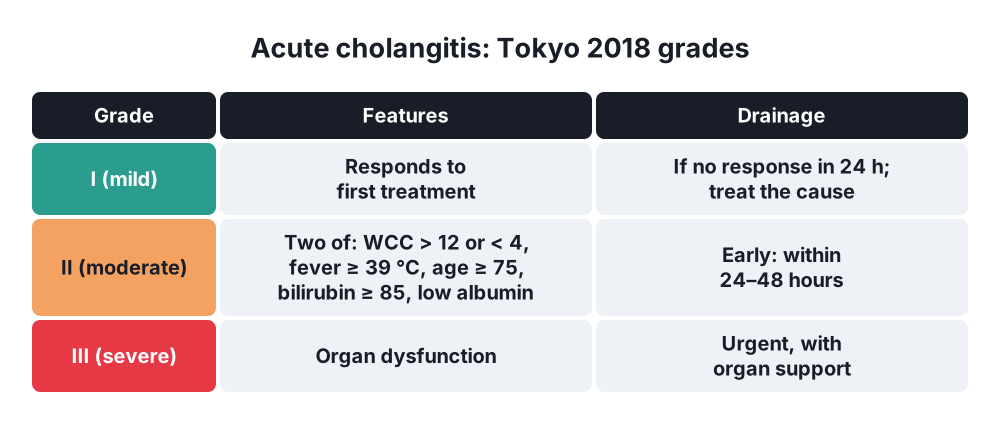
Tokyo Guidelines 2018: severity
- Grade I (mild) → responds to initial treatment
- Grade II (moderate) → any two of:
- White cell count over 12 or under 4 × 10⁹/L
- Fever ≥ 39 °C
- Age ≥ 75
- Bilirubin ≥ 85 µmol/L (5 mg/dL)
- Albumin under 0.7 × lower limit
- Grade III (severe) → organ dysfunction (cardiovascular, neurological, respiratory, renal, hepatic, haematological)
Other
- Acute, recurrent, suppurative
- Recurrent pyogenic cholangitis
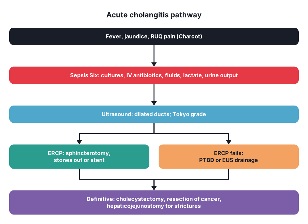
- Charcot's triad and Reynolds' pentad (clinical)
6. Clinical picture
Symptoms
- Charcot's triad (about 50–70%):
- Fever with rigors
- Jaundice
- RUQ pain
- Reynolds' pentad (severe, suppurative; about 5–7%) → Charcot's triad plus:
- Hypotension (shock)
- Confusion
- Dark urine, pale stools, itch
- Elderly → may present with confusion, falls or hypotension only
- History → gallstones, biliary surgery, stent, ERCP
Signs
- High swinging fever, rigors
- Jaundice
- RUQ tenderness; usually no peritonism
- Septic shock → tachycardia, hypotension, oliguria, altered mental state
- Liver may be tender and enlarged
7. Diagnosis
- Bloods:
- FBC → leucocytosis (or low white count in severe sepsis)
- CRP
- LFT → raised bilirubin, ALP, GGT (cholestatic); transaminases may rise sharply early with a stone
- U&E, creatinine, coagulation (INR), platelets, albumin
- Blood cultures (positive in 20–70%), lactate, blood gas
- Amylase or lipase (gallstone pancreatitis)
- Ultrasound → first test: dilated CBD (over 6–7 mm) and intrahepatic ducts, stones, liver abscess
- CT → cause (tumour, stricture, stent), abscess; dilated ducts
- MRCP → stones and strictures; plan therapy in stable patients
- EUS → small stones, ampullary tumours
- ERCP → diagnostic and therapeutic; bile culture
- Score sepsis → qSOFA, SOFA; Tokyo grade
8. Differential diagnosis
- Acute cholecystitis (no or mild jaundice, Murphy's sign)
- Gallstone pancreatitis (often together)
- Liver abscess
- Viral or alcoholic hepatitis
- Right lower lobe pneumonia, pyelonephritis (fever and pain)
- Primary sclerosing cholangitis (chronic, ulcerative colitis, beaded ducts)
- IgG4-related sclerosing cholangitis
- Sepsis from another source with cholestasis
9. Treatment
Conservative
- Sepsis Six within the first hour:
- Oxygen
- Blood cultures
- IV antibiotics → broad-spectrum, e.g. piperacillin-tazobactam; or ceftriaxone with metronidazole; meropenem for severe or resistant cases (local guidance)
- IV fluids (balanced crystalloid)
- Lactate
- Urine output (catheter)
- Correct coagulopathy (vitamin K, FFP or platelets before procedures)
- Critical care → vasopressors, organ support for grade III
- Antibiotics duration → 4–7 days after source control; 2 weeks if Enterococcus or streptococcal bacteraemia
- Analgesia
Surgical: biliary drainage
- Timing by Tokyo grade:
- Grade I → antibiotics; drainage if no response in 24 hours; treat the cause (ERCP, cholecystectomy)
- Grade II → early drainage within 24–48 hours
- Grade III → urgent drainage as soon as organ support starts (within 12–24 hours)
- ERCP → first choice:
- Endoscopic sphincterotomy with stone extraction, or
- Plastic stent or nasobiliary drain alone in the very sick (quick decompression; stones cleared later)
- Coagulopathy → stent without sphincterotomy
- Percutaneous transhepatic biliary drainage (PTBD) → ERCP failed or not possible (altered anatomy after gastric bypass or hepaticojejunostomy), hilar strictures
- EUS-guided biliary drainage (choledochoduodenostomy, hepaticogastrostomy) → failed ERCP in expert centres
- Open surgical drainage → rarely now; when endoscopic and percutaneous routes fail:
- CBD exploration with T-tube drainage
- Mortality much higher than endoscopic drainage in sick patients
- Definitive treatment of the cause once recovered:
- Laparoscopic cholecystectomy after stone cholangitis (same admission or within weeks)
- Cancer → staging and resection (Whipple, hepatectomy) or metal stent (palliation)
- Benign strictures → endoscopic stents, Roux-en-Y hepaticojejunostomy
- Recurrent pyogenic cholangitis → clear stones, hepatectomy of the affected segments, hepaticojejunostomy with access loop
10. Complications
- Septic shock, multiorgan failure, death
- Liver abscesses (multiple, pyogenic)
- Portal vein thrombosis (pylephlebitis)
- Acute kidney injury (sepsis, jaundice: hepatorenal)
- Recurrent cholangitis, secondary biliary cirrhosis (long-standing obstruction)
- ERCP complications → post-ERCP pancreatitis, bleeding after sphincterotomy, duodenal perforation, stent blockage or migration
- PTBD → bleeding (haemobilia), bile leak, catheter dislodgement
Prognosis
- Poor outcome → shock, confusion, renal failure, age over 75, malignant obstruction, delayed drainage
- Mortality → grade I under 2%; grade III about 5–10%
Show as a list
Acute cholangitis
- Cause
- Obstruction + infection
- CBD stones
- Strictures, cancer
- Blocked stent
- Bugs
- E. coli
- Klebsiella
- Enterococcus
- Clinical
- Charcot's triad
- Reynolds' pentad
- Tests
- LFT cholestatic
- Blood cultures
- Ultrasound: dilated CBD
- MRCP
- Tokyo grades
- I: mild
- II: drain 24-48 h
- III: urgent drain
- Treatment
- Sepsis Six
- Antibiotics
- ERCP: stone out or stent
- PTBD if ERCP fails
- Lap chole later


