33. Chest injuries, pneumothorax and haemothorax
High yield
Blunt and penetrating chest trauma; tension and open pneumothorax, haemothorax, flail chest and tamponade found in the primary survey.
Exam Prep · State examSurgery · Emergency and traumaAnatomy · Thorax
1. Definition
- Chest injury → trauma to the chest wall, pleura, lungs, heart, great vessels, oesophagus or diaphragm
- Pneumothorax → air in the pleural cavity
- Haemothorax → blood in the pleural cavity
- Chest injuries → about 25% of trauma deaths
- 50% of patients dying of multiple injuries have a significant chest injury
- Most treated without thoracotomy → chest drain alone in about 85%
2. Aetiology and risk factors
- Blunt → road traffic accidents (most common), falls, crush, blast, deceleration
- Penetrating → stab, gunshot
- Iatrogenic → central line insertion, lung biopsy, positive pressure ventilation
3. Pathogenesis
- Loss of negative intrapleural pressure → lung collapse
- Tension: one-way valve → air enters on inspiration, cannot leave
- Rising pressure in the hemithorax
- Mediastinal shift to the opposite side
- Kinking of the venae cavae → reduced venous return
- Low cardiac output → obstructive shock, cardiac arrest
- Open pneumothorax → wound > 2/3 of the tracheal diameter; air goes through the wound instead of the trachea
- Flail chest → paradoxical movement of a free segment; hypoxia mainly from the underlying pulmonary contusion
- Haemothorax → bleeding from intercostal or internal thoracic arteries, lung, heart, great vessels
4. Classification
By mechanism
- Blunt
- Penetrating
By structure
- Chest wall → rib fractures, flail chest, sternal fracture
- Pleura → pneumothorax, haemothorax
- Lung → contusion, laceration
- Heart → tamponade, blunt cardiac injury
- Great vessels → traumatic aortic rupture
- Airway → tracheobronchial rupture
- Oesophagus, diaphragm
Pneumothorax types
- Simple (closed)
- Tension
- Open (sucking chest wound)
Haemothorax by volume on chest X-ray
- Small → < 500 mL, blunting of the costophrenic angle
- Medium → 500–1500 mL
- Massive → > 1500 mL
Immediately life-threatening (primary survey)
- Airway obstruction
- Tension pneumothorax
- Open pneumothorax
- Massive haemothorax
- Flail chest with pulmonary contusion
- Cardiac tamponade
5. Clinical picture
Symptoms
- Chest pain, worse on breathing
- Dyspnoea
- Haemoptysis
Signs
- Tachypnoea, cyanosis, low oxygen saturation
- Bruising, seat belt sign, wounds
- Subcutaneous emphysema → crepitus under the skin
- Simple pneumothorax → reduced breath sounds, hyperresonance
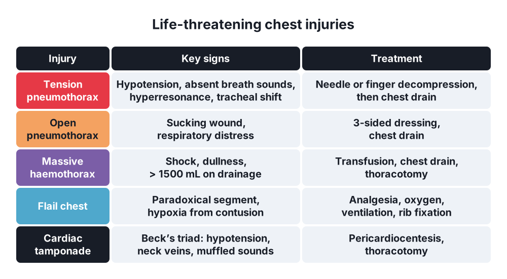
- Tension pneumothorax:
- Respiratory distress, tachycardia, hypotension
- Absent breath sounds, hyperresonance on the affected side
- Tracheal deviation to the opposite side (late sign)
- Distended neck veins
- Haemothorax → reduced breath sounds, dullness on percussion, shock
- Flail chest → paradoxical segment, crepitus of ribs
- Cardiac tamponade → Beck's triad: hypotension, distended neck veins, muffled heart sounds; pulsus paradoxus
- Traumatic aortic rupture → deceleration injury, unequal arm pulses, widened mediastinum
- Diaphragmatic rupture → blunt seat belt injury, mostly left side; raised left hemidiaphragm or bowel in the chest
- Tracheobronchial rupture → persistent large air leak after chest drain
6. Diagnosis
- Tension pneumothorax → clinical diagnosis; treat before any X-ray
Imaging
- Extended focused assessment with sonography for trauma (eFAST):
- Pneumothorax → absent lung sliding; barcode sign on M-mode
- Haemothorax, pericardial fluid
- More sensitive than chest X-ray for pneumothorax
- Chest X-ray:
- Visceral pleural line, no lung markings beyond → pneumothorax
- Deep sulcus sign → pneumothorax on a supine film
- Haemothorax → diffuse haziness on a supine film; meniscus when erect
- Widened mediastinum > 8 cm → aortic injury
- CT chest with contrast → stable patient; occult pneumothorax, contusion, aorta
- Electrocardiogram (ECG), troponin → blunt cardiac injury
- Bronchoscopy → tracheobronchial injury
- Oesophagoscopy or contrast swallow → oesophageal injury
Laboratory
- Arterial blood gas
- Full blood count, cross-match
7. Differential diagnosis
- Spontaneous pneumothorax
- Pleural effusion
- Lung collapse, endobronchial intubation
- Diaphragmatic rupture with bowel in the chest
- Pulmonary embolism, myocardial infarction
8. Treatment
Primary survey (ATLS)
- High-flow oxygen, cervical spine control
- Airway → intubate if needed
- Tension pneumothorax → immediate decompression:
- Needle decompression with a 12G cannula → 5th intercostal space between anterior and mid-axillary lines (current ATLS, adults), or 2nd intercostal space, midclavicular line (classic site)
- Finger thoracostomy → 5th intercostal space, same position
- Always followed by a chest drain in the 5th intercostal space
- Open pneumothorax → 3-sided occlusive dressing, then chest drain away from the wound and closure of the wound
- Massive haemothorax → 2 large-bore cannulas, blood transfusion, chest drain
- Cardiac tamponade → pericardiocentesis (ultrasound-guided) or thoracotomy
Chest drain (tube thoracostomy)
- Indications (British Thoracic Society):
- Pneumothorax in a ventilated patient
- Tension pneumothorax after needle decompression
- Persistent or recurrent pneumothorax after aspiration
- Traumatic haemopneumothorax
- Simple traumatic pneumothorax that is large, symptomatic, or before general anaesthesia
- Empyema, malignant effusion, after thoracic surgery
- Patient at 45°, arm abducted; pulse oximetry
- Triangle of safety → anterior border of latissimus dorsi, lateral border of pectoralis major, line of the 5th intercostal space (nipple level), apex below the axilla
- Local anaesthetic down to the parietal pleura
- 2 cm incision; blunt dissection over the upper border of the rib below → avoids the neurovascular bundle
- Pierce the pleura with a blunt clamp; finger sweep for adhesions
- Insert the tube without a trocar → upwards for air, downwards for blood
- Underwater seal; secure with suture; check swing and bubbling; chest X-ray
- Smaller tubes (12–20 Fr) are effective; wide-bore drain for massive haemothorax
- Too low or too posterior → injury to liver, spleen, diaphragm
Indications for thoracotomy
- Initial drainage > 1500 mL of blood
- Continued drainage > 200 mL/h for 2–4 h
- Need for repeated transfusion
- Cardiac tamponade
- Large air leak → tracheobronchial injury
- Oesophageal, great vessel or diaphragm injury
- Emergency department (resuscitative) thoracotomy → penetrating chest trauma with arrest in the department
Other injuries
- Simple pneumothorax → chest drain; small and stable → observe
- Retained haemothorax → video-assisted thoracoscopic surgery (VATS) within 3–7 days
- Rib fractures, flail chest:
- Small segment, adequate breathing → high-dependency unit, analgesia, physiotherapy, early mobilisation, serial blood gases
- Analgesia → epidural, paravertebral or serratus anterior block
- Severe → intubation and positive pressure ventilation
- Surgical rib fixation → selected flail chest
- Pulmonary contusion → most common chest injury that can become lethal; oxygen, analgesia, physiotherapy, avoid fluid overload, ventilation if hypoxic
- Tracheobronchial, aortic, cardiac or diaphragmatic injury → CT, cardiothoracic referral
- Traumatic aortic rupture → blood pressure control, endovascular stent graft
9. Complications
- Empyema → infected retained haemothorax
- Fibrothorax, trapped lung → decortication
- Pneumonia, acute respiratory distress syndrome (ARDS)
- Persistent air leak, bronchopleural fistula
- Re-expansion pulmonary oedema
- Chest drain → malposition, organ injury, bleeding from intercostal vessels
- Chronic chest wall pain
Show as a list
Chest injuries
- Mechanism
- Blunt
- Penetrating
- Pneumothorax
- Simple
- Tension
- Open
- Haemothorax
- Small, medium
- Massive > 1500 mL
- Other
- Flail chest
- Tamponade
- Aortic rupture
- Diagnosis
- Clinical
- eFAST
- Chest X-ray, CT
- Treatment
- Decompression
- Chest drain
- Thoracotomy


