
61. Bleeding peptic ulcer
High yield
Commonest cause of upper gastrointestinal bleeding; endoscopic dual therapy and PPI first; surgery or embolisation if endoscopy fails.
Exam Prep · State examSurgery · Upper GISurgery · Emergency and trauma
1. Definition
- Bleeding peptic ulcer → haemorrhage from a gastric or duodenal ulcer eroding a vessel in its base
- Upper gastrointestinal (UGI) bleeding → source proximal to the ligament of Treitz
2. Epidemiology
- Peptic ulcer → 36% of UGI bleeding (commonest cause)
- Mortality of ulcer bleeding → 5.8%; after surgery → 30%
3. Aetiology and risk factors
- Helicobacter pylori (H. pylori)
- Non-steroidal anti-inflammatory drugs (NSAIDs), aspirin
- Antiplatelet and anticoagulant drugs
- Elderly patients with comorbidity
- Stress ulcers → intensive care, burns, head injury
4. Pathogenesis
- Ulcer base erodes an artery:
- Posterior duodenal ulcer → gastroduodenal artery (GDA)
- Lesser curvature gastric ulcer → left gastric artery
- Anterior duodenal ulcers → ooze or perforate, rarely bleed massively
- Bleeding gastric ulcers → antrum or incisura (77%), higher lesser curve (15%)
- Acid (pH < 6) → impairs platelet aggregation and clot stability
5. Classification
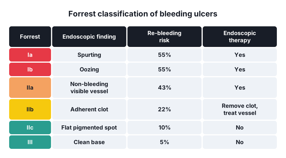
Forrest classification (endoscopic)
- Ia → spurting arterial bleeding
- Ib → oozing
- IIa → non-bleeding visible vessel
- IIb → adherent clot
- IIc → flat pigmented spot
- III → clean base
Risk of further bleeding (without endoscopic therapy)
- Active bleeding → 55%
- Visible vessel → 43%
- Adherent clot → 22%
- Flat spot → 10%
- Clean base → 5%
Risk scores
- Glasgow-Blatchford score (GBS) → before endoscopy; predicts need for intervention; GBS 0 → safe discharge
- Rockall score:
- Pre-endoscopy → age, shock, comorbidity
- Full → adds diagnosis and stigmata of recent haemorrhage
- Predicts mortality better than re-bleeding
6. Clinical picture
Symptoms
- Haematemesis → fresh blood; worse prognosis
- Coffee-ground vomiting → altered blood; not active bleeding
- Melaena → black tarry stool
- Haematochezia → massive UGI bleeding
- Syncope, dizziness
- History of dyspepsia, NSAID or anticoagulant use
Signs
- Tachycardia, hypotension, postural drop
- Pallor, cold peripheries
- Melaena on rectal examination
- Epigastric tenderness
7. Diagnosis
Laboratory
- Haemoglobin → poor guide to acute blood loss
- Urea → raised (digested blood)
- Clotting screen, platelets
- Crossmatch
- Liver function tests → suspected varices
Imaging
- Oesophagogastroduodenoscopy (OGD) → diagnosis and treatment
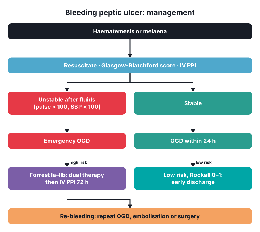
- Unstable or ongoing bleeding → emergency OGD after resuscitation
- Stable → within 24 h
- CT angiography → bleeding not found or not controlled at endoscopy
- Mesenteric angiography → localisation and embolisation
8. Differential diagnosis
- Oesophageal or gastric varices
- Mallory-Weiss tear
- Oesophagitis
- Gastritis, erosive duodenitis
- UGI malignancy
- Dieulafoy's lesion → within 6 cm of the cardia
- Aortoduodenal fistula → previous aortic surgery
9. Treatment
Conservative
- Resuscitation:
- 2 large-bore cannulas
- Crystalloid
- Red cells when ≥ 30% blood volume lost
- Platelets → keep > 50 × 10⁹/L
- Fresh frozen plasma (FFP) → after > 1 blood volume lost
- Intravenous proton pump inhibitor (PPI) before endoscopy
- Endoscopic therapy → Forrest Ia, Ib, IIa; IIb after clot removal:
- Dual therapy → adrenaline 1:10 000 injection + clip or thermal coagulation
- Adrenaline alone → not adequate
- Haemostatic powder → rescue
- After endoscopic therapy → high-dose PPI: omeprazole 80 mg bolus, then 8 mg/h for 72 h
- H. pylori → test, eradicate, confirm eradication
- Re-bleeding:
- Repeat endoscopic therapy → controls 75%
- Transarterial embolisation (TAE) → controls 64–91%; first choice after failed surgery
- Surgery → ulcer > 2 cm with shock
- Endoscopic failure more likely:
- Ulcer > 2 cm
- Lesser curvature ulcer
- Posterior or superior wall of the duodenal bulb
Surgical
- Indications:
- Massive bleeding not controlled at endoscopy
- Re-bleeding after 2 endoscopic attempts
- Re-bleeding with shock from an ulcer > 2 cm
- TAE not available or failed
- Bleeding duodenal ulcer:
- Kocher manoeuvre → mobilise the duodenum
- Longitudinal duodenotomy across the pylorus
- Control bleeding with finger pressure
- Under-run the GDA above and below the ulcer (0 or 1-0 suture)
- Medial U-stitch → transverse pancreatic artery
- No lateral stitch → common bile duct (CBD)
- Close as a Heineke-Mikulicz pyloroplasty
- Bleeding gastric ulcer:
- Under-run or excise the ulcer, with biopsy
- Distal gastrectomy → large or suspicious ulcer
- Vagotomy → no longer added; PPI and eradication replace it
- Giant posterior DU → distal gastrectomy with ulcer exclusion, controlled duodenostomy
10. Complications
- Re-bleeding
- Duodenal leak or stump blow-out
- CBD injury from deep stitches
- Pancreatitis
- Aspiration pneumonia
- Multi-organ failure
- Ischaemic events → myocardial infarction, stroke
11. Prognosis
- Mortality → 5.8% for all patients; 30% after surgery
- Rockall score → predicts death
Show as a list
Bleeding peptic ulcer
- Causes
- H. pylori
- NSAIDs
- Antiplatelets
- Vessels
- Posterior DU: GDA
- Lesser curve GU: left gastric
- Assessment
- Glasgow-Blatchford
- Rockall
- Forrest
- Resuscitation
- Large-bore cannulas
- Blood products
- IV PPI
- Endoscopy
- Dual therapy
- Clips
- Thermal probe
- Re-bleeding
- Repeat endoscopy
- Embolisation
- Surgery
- Surgery
- Duodenotomy
- Under-run GDA
- Pyloroplasty closure