
48. Atherosclerotic and diabetic gangrene of the limbs
High yield
Dry or wet tissue death from arterial occlusion; diabetic foot combines neuropathy, ischaemia and infection; debride, revascularise, amputate.
Exam Prep · State examSurgery · General surgerySurgery · Emergency and trauma
1. Definition
- Gangrene → death of a large area of tissue, with putrefaction
- Atherosclerotic gangrene → tissue death from end-stage arterial occlusion (Fontaine IV, CLTI)
- Diabetic gangrene (diabetic foot) → ulceration, infection or gangrene of the foot in a diabetic, from neuropathy, ischaemia and infection
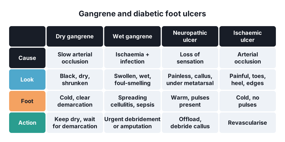
- Dry gangrene → dry, shrunken, mummified tissue without infection
- Wet gangrene → moist, swollen, infected tissue with spreading putrefaction
2. Epidemiology
- Diabetic foot ulcer → lifetime risk up to 25% in diabetes
- Diabetics are about 15 times more likely to have a major amputation
- 85% of diabetic amputations start with an ulcer
- Diabetic ulcers → 45% neuropathic, 10% ischaemic, 45% mixed neuro-ischaemic
- 5-year mortality after major amputation → about 50–70%
3. Aetiology and risk factors
Atherosclerotic gangrene
- Progressive atherosclerosis → smoking, hypertension, hyperlipidaemia, age
- Acute thrombosis on a plaque, embolism
- Minor trauma on an ischaemic foot (tight shoes, nail cutting)
Diabetic foot
- Neuropathy:
- Sensory → painless injury not noticed
- Motor → clawed toes, high pressure under metatarsal heads
- Autonomic → dry, cracked skin; arteriovenous shunting
- Ischaemia → diffuse, distal (tibial) disease; calcified arteries (Mönckeberg's sclerosis)
- Infection → poor immunity from high glucose
- Previous ulcer or amputation, deformity, Charcot foot, callus
- Poor glucose control, kidney failure, poor vision, living alone
- Organisms → S. aureus, streptococci, Gram-negatives, anaerobes (often mixed)
4. Pathogenesis
Dry gangrene
- Slow fall in arterial flow
- Tissue dies and dries out → coagulative necrosis
- Haemoglobin breaks down → black colour (iron sulphide)
- Line of demarcation forms → band of granulation tissue between dead and living tissue
- Dead part may separate on its own (autoamputation)
Wet gangrene
- Fast ischaemia, venous congestion and infection
- Liquefactive necrosis, putrefaction by bacteria
- No clear demarcation → spreads quickly
- Toxins absorbed → sepsis
- Gas gangrene → Clostridium; crepitus; surgical emergency
Diabetic foot
- Neuropathy → unnoticed injury → ulcer
- Ischaemia → poor healing
- Infection → spreads along tendon sheaths and plantar spaces → abscess, osteomyelitis, wet gangrene
- Charcot foot → neuropathic joint destruction; "rocker-bottom" deformity
5. Classification
By type
- Dry → atherosclerotic, embolic, Buerger's
- Wet → diabetic, venous obstruction, infected dry gangrene
- Gas gangrene (Clostridium)
Wagner (diabetic foot)
- 0 → no ulcer, foot at risk (deformity, callus)
- 1 → superficial ulcer
- 2 → deep ulcer to tendon, capsule or bone
- 3 → deep ulcer with abscess or osteomyelitis
- 4 → forefoot gangrene
- 5 → whole-foot gangrene
Others
- University of Texas → depth plus infection and ischaemia
- WIfI → wound, ischaemia, foot infection
6. Clinical picture
Symptoms
- Atherosclerotic → claudication, then rest pain, then black toes
- Diabetic neuropathic → painless ulcer, numb "stocking" feet
- Wet gangrene → swelling, smell, discharge, fever, confusion (sepsis)
Signs
- Dry gangrene:
- Black, dry, shrunken, cold
- Clear line of demarcation
- No pulse, no sensation, no capillary refill
- Wet gangrene:
- Swollen, blistered, discoloured, foul-smelling
- Cellulitis spreading up the leg, lymphangitis
- Crepitus → gas-forming organisms
- Neuropathic ulcer → warm foot, palpable pulses, painless, under the metatarsal head or heel, surrounded by callus
- Ischaemic ulcer → cold foot, absent pulses, painful, on toes, heel, edges of the foot
- Probe-to-bone test → positive suggests osteomyelitis
- Sensation → 10 g monofilament, vibration (128 Hz tuning fork)
7. Diagnosis
Laboratory
- FBC, CRP, ESR, glucose, HbA1c, U&E (kidney function), lactate
- Blood cultures, deep tissue or bone culture (not surface swabs)
Vascular assessment
- Pulses, ABPI → falsely high in diabetics (calcification)
- Toe pressures, toe-brachial index, transcutaneous oxygen (TcPO₂)
- Duplex ultrasound, CT or MR angiography, digital subtraction angiography → plan revascularisation
Imaging of the foot
- Plain X-ray → gas in tissues, osteomyelitis, Charcot changes, foreign body
- MRI → osteomyelitis, abscess, extent of infection
- Bone scan, white cell scan
8. Differential diagnosis
- Neuropathic versus ischaemic versus venous ulcer
- Necrotising fasciitis
- Cellulitis without gangrene
- Charcot foot versus osteomyelitis
- Frostbite, Buerger's disease, vasculitis
- Embolic "blue toe" syndrome
9. Treatment
- Team → multidisciplinary diabetic foot team: vascular surgeon, diabetologist, podiatrist, orthotist, microbiologist, nurses
Conservative
- Prevention:
- Daily foot inspection, proper footwear, never walk barefoot
- Podiatry → callus and nail care
- Annual foot screening
- Glucose control, stop smoking, statin, antiplatelet
- Offloading → total contact cast, removable boot, bed rest
- Antibiotics → broad-spectrum IV for severe infection, then culture-guided; 6 weeks for osteomyelitis
- Wound care → moist dressings, negative pressure therapy
- Dry gangrene → keep dry and cool; never heat the limb; wait for demarcation if no infection
Surgical
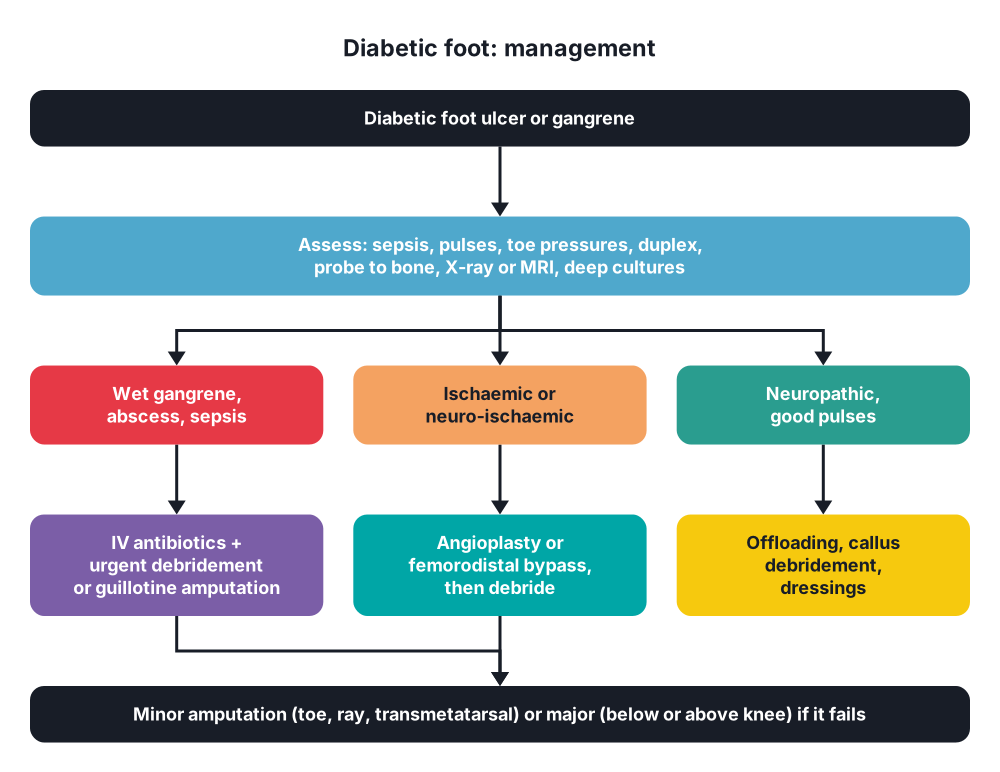
- Wet gangrene or abscess → urgent debridement and drainage; open all infected spaces; remove dead tissue and bone
- Revascularisation → for ischaemic or mixed ulcers:
- Angioplasty (tibial arteries)
- Femorodistal bypass with vein
- Do it before or soon after debridement, so wounds can heal
- Amputation → at the lowest level that will heal:
- Toe, ray, transmetatarsal → diabetic forefoot disease
- Below-knee (long posterior or skew flap; tibia cut 10–15 cm below the knee joint) → best for walking with a prosthesis
- Above-knee → non-ambulant, failed below-knee, poor blood flow at the knee
- Guillotine amputation → life-threatening sepsis; formal stump 2–5 days later
- Wound cover → skin graft, flaps
10. Complications
- Sepsis, death
- Osteomyelitis
- Major amputation, amputation of the other leg (50% within 5 years)
- Stump problems → infection, failure to heal, re-amputation, phantom limb pain, contracture
- Cardiovascular death
- Recurrence of ulcers (up to 40% in 1 year)
Show as a list
Gangrene of the limbs
- Dry
- Atherosclerosis
- Coagulative necrosis
- Demarcation line
- Wet
- Infection
- Liquefactive necrosis
- Sepsis
- Diabetic foot
- Neuropathy
- Ischaemia
- Infection
- Wagner 0-5
- Diagnosis
- Toe pressures
- Duplex, angiography
- X-ray, MRI
- Treatment
- Offloading
- Antibiotics
- Debridement
- Revascularisation
- Amputation level


