
75. Acute pancreatitis
High yield
Gallstones and alcohol cause most; diagnose by Atlanta criteria, grade severity, treat supportively, and step up for infected necrosis.
Exam Prep · State examSurgery · HPBSurgery · Emergency and trauma
1. Definition
- Acute pancreatitis → acute inflammation of the pancreas, from premature activation of enzymes inside the gland, with variable involvement of nearby tissues and distant organs
- Diagnosis (Revised Atlanta 2012) → two of three:
- Typical pain → acute, severe, persistent epigastric pain, often radiating to the back
- Serum lipase or amylase ≥ 3 times the upper limit of normal
- Typical imaging findings (CT, MRI or ultrasound)
2. Epidemiology
- About 30–40 per 100,000 a year; rising
- Causes → gallstones about 40–50%, alcohol about 20–30% (gallstones more in women, alcohol more in men)
- Mild disease about 80% → recovers in days; mortality under 1%
- Severe disease about 20% → persistent organ failure; mortality 15–30%
- Infected necrosis → mortality up to 30%
- Two peaks of death → early (first week) from organ failure (SIRS); late (after 2 weeks) from infected necrosis and sepsis
3. Aetiology and risk factors
Causes ("I GET SMASHED")
- Idiopathic → 10–20% (many are microlithiasis)
- Gallstones → most common; small stones (under 5 mm) pass and block the ampulla
- Ethanol (alcohol)
- Trauma (blunt abdominal injury)
- Steroids
- Mumps and other viruses (Coxsackie, CMV, HIV), Ascaris
- Autoimmune (IgG4-related pancreatitis)
- Scorpion sting (Tityus trinitatis)
- Hypertriglyceridaemia (over 11.3 mmol/L), Hypercalcaemia (hyperparathyroidism), Hypothermia
- ERCP (post-ERCP pancreatitis 3–5%; most common iatrogenic cause)
- Drugs → azathioprine, valproate, thiazides, furosemide, oestrogens, didanosine, L-asparaginase, GLP-1 agonists (reported)
Other
- Pancreas divisum, tumours (IPMN, pancreatic cancer in older patients), sphincter of Oddi dysfunction
- Genetic → PRSS1, SPINK1, CFTR mutations
- Ischaemia (after cardiac surgery), pregnancy, smoking
4. Pathogenesis
- Trigger → duct obstruction (stone), alcohol, toxins
- Trypsinogen activated to trypsin inside the acinar cell (fusion of lysosomes and zymogen granules, calcium overload)
- Trypsin activates other enzymes → elastase, phospholipase A2, lipase → autodigestion
- Local injury → oedema, fat necrosis, haemorrhage, necrosis of the gland
- Cytokine release (IL-1, IL-6, TNF-alpha) → systemic inflammatory response (SIRS)
- Capillary leak, third-space fluid loss, hypovolaemia
- Organ failure → lungs (ARDS), kidneys, circulation
- Gut barrier failure → bacterial translocation → infected necrosis (second to fourth week)
- Fat necrosis binds calcium → hypocalcaemia
- Islet damage → hyperglycaemia
5. Classification
Revised Atlanta: types
- Interstitial oedematous pancreatitis (about 85–90%) → enhancing gland with oedema
- Necrotising pancreatitis (5–10%) → necrosis of the gland, the fat around it, or both
Revised Atlanta: severity
- Mild → no organ failure, no local or systemic complications
- Moderately severe → transient organ failure (under 48 hours) and/or local complications
- Severe → persistent organ failure (over 48 hours), single or multiple
- Organ failure → modified Marshall score ≥ 2 in respiratory, renal or cardiovascular systems
Local complications (Revised Atlanta)
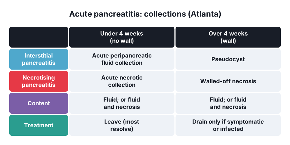
- Under 4 weeks:
- Acute peripancreatic fluid collection (APFC) → interstitial pancreatitis; no wall
- Acute necrotic collection (ANC) → necrotising pancreatitis; solid and liquid, no wall
- Over 4 weeks (wall formed):
- Pseudocyst → fluid only, well-defined wall (interstitial pancreatitis)
- Walled-off necrosis (WON) → solid and liquid necrosis with a wall
- Any of these can be sterile or infected
- Other → gastric outlet obstruction, splenic or portal vein thrombosis, colonic necrosis
Severity scores
- Glasgow (Imrie) at 48 hours → 3 or more = severe ("PANCREAS"):
- PaO₂ < 8 kPa
- Age > 55
- Neutrophils (WCC) > 15 × 10⁹/L
- Calcium < 2 mmol/L
- Renal: urea > 16 mmol/L
- Enzymes: LDH > 600 IU/L, AST > 200 IU/L
- Albumin < 32 g/L
- Sugar: glucose > 10 mmol/L
- Ranson's (on admission and at 48 hours), APACHE II (≥ 8 = severe), BISAP
- CRP > 150 mg/L at 48 hours → severe disease
- CT severity index (Balthazar) → grade of inflammation plus extent of necrosis
- Persistent SIRS over 48 hours → predicts organ failure and death
6. Clinical picture
Symptoms
- Sudden severe constant epigastric pain radiating to the back; better sitting forward
- Nausea and vomiting (repeated)
- After a heavy meal or alcohol binge; history of gallstones
- Breathlessness, confusion in severe cases
Signs
- Tachycardia, fever, tachypnoea, hypotension
- Epigastric tenderness and guarding (often less than the pain suggests), distension, ileus
- Jaundice → stone in the CBD, cholangitis
- Grey Turner's sign (flank bruising) and Cullen's sign (periumbilical bruising) → retroperitoneal bleeding in severe disease (late, 1–3%)
- Fox's sign (bruising over the inguinal ligament)
- Pleural effusion (left), basal crackles
- Hypocalcaemia → Chvostek's and Trousseau's signs
- Xanthelasma (hyperlipidaemia), signs of chronic alcohol use
7. Diagnosis
Blood tests
- Lipase (more specific, stays high longer) or amylase ≥ 3 × normal
- Amylase may be normal in hypertriglyceridaemia and late presentation; high in perforated ulcer, mesenteric ischaemia, ectopic pregnancy, parotitis
- FBC, CRP, U&E (urea, creatinine), LFT (ALT over 3 × normal suggests gallstones), calcium, glucose, triglycerides, LDH, albumin, arterial blood gas (PaO₂, lactate)
Imaging
- Ultrasound within 24 hours → gallstones (look in every case), CBD dilatation
- Contrast CT → not routine on admission:
- Only if the diagnosis is uncertain
- Best at 72–96 hours to assess necrosis (early CT underestimates it)
- Later for deterioration and collections
- MRCP or EUS → CBD stones, microlithiasis, idiopathic cases
- Chest X-ray → effusion, ARDS; erect film to exclude perforation
- Abdominal X-ray → sentinel loop, colon cut-off sign
Cause in idiopathic cases
- Repeat ultrasound, EUS (microlithiasis, small tumours), MRCP (divisum), calcium, triglycerides, IgG4; genetic testing in young patients; CT in over-40s (pancreatic cancer)
8. Differential diagnosis
- Perforated peptic ulcer (free air, sudden onset)
- Acute cholecystitis, biliary colic, cholangitis
- Mesenteric ischaemia (amylase raised, lactate high)
- Ruptured abdominal aortic aneurysm
- Myocardial infarction (inferior)
- Small bowel obstruction
- Diabetic ketoacidosis
- Ectopic pregnancy
9. Treatment
Conservative
- Supportive care → no specific drug treats acute pancreatitis
- Assess severity → admit; HDU or ICU for organ failure or persistent SIRS
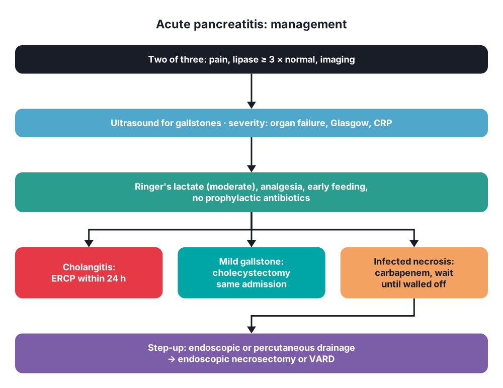
- IV fluids → goal-directed, moderate with Ringer's lactate (WATERFALL trial: aggressive fluid resuscitation increases fluid overload without benefit)
- About 1.5 mL/kg/h after a bolus in hypovolaemia; target urine output > 0.5 mL/kg/h, falling haematocrit and urea
- Analgesia → opioids (morphine is safe), paracetamol; epidural in severe pain
- Oxygen; monitor saturations, blood gases
- Early oral feeding in mild disease, as soon as tolerated (within 24 hours); no need to wait for enzymes to fall
- Enteral nutrition within 24–72 hours in severe disease → nasogastric or nasojejunal (equal); better than parenteral (fewer infections, less organ failure, lower mortality)
- No prophylactic antibiotics → even in necrotising disease; give antibiotics only for proven or suspected infection (cholangitis, infected necrosis, pneumonia)
- VTE prophylaxis; catheter, monitoring of glucose and calcium
- Treat the cause → stop alcohol and offending drugs; insulin and plasmapheresis for severe hypertriglyceridaemia; treat hypercalcaemia
Surgical: gallstone pancreatitis
- Urgent ERCP (within 24 hours) → cholangitis only
- ERCP within 72 hours → persistent biliary obstruction (rising bilirubin, dilated CBD)
- No routine early ERCP for gallstone pancreatitis without cholangitis (APEC trial)
- Cholecystectomy:
- Mild → same admission (within 2 weeks; PONCHO trial: fewer readmissions)
- Severe or with collections → delay until collections resolve or for 6 weeks
- Unfit → ERCP with sphincterotomy to prevent recurrence
Surgical: necrotising pancreatitis
- Sterile necrosis → conservative (no intervention), even if extensive
- Infected necrosis → suspect with fever, rising CRP, deterioration after 7–10 days; gas in the collection on CT; FNA rarely needed
- Antibiotics that penetrate necrosis → carbapenems (meropenem), quinolones with metronidazole
- Delay intervention until the necrosis is walled off (ideally after 4 weeks) → safer, less bleeding
- Step-up approach (PANTER, TENSION trials):
- Drain first → endoscopic transgastric drainage (EUS-guided lumen-apposing metal stent) or percutaneous retroperitoneal drainage
- Then, if no improvement → endoscopic necrosectomy or video-assisted retroperitoneal debridement (VARD)
- Open necrosectomy → rarely now; when minimally invasive methods fail, or for complications (bleeding, bowel ischaemia, perforation)
- About one-third of infected necrosis recovers with drainage alone
- Pseudocyst → drain only if symptomatic (pain, obstruction, infection, bleeding) or growing; endoscopic cystgastrostomy preferred (see question 76)
- Abdominal compartment syndrome → bladder pressure over 20 mmHg with new organ failure; medical measures, percutaneous drainage of ascites, decompressive laparotomy if refractory
- Bleeding pseudoaneurysm → angioembolisation
10. Complications
Systemic
- SIRS and organ failure → ARDS, acute kidney injury, shock
- Disseminated intravascular coagulation
- Hypocalcaemia, hyperglycaemia
- Abdominal compartment syndrome
Local
- Acute peripancreatic fluid collection, acute necrotic collection
- Pseudocyst, walled-off necrosis
- Infected necrosis, pancreatic abscess
- Pseudoaneurysm (splenic, gastroduodenal artery) → bleeding
- Splenic or portal vein thrombosis → gastric varices
- Pancreatic ascites, pancreatic fistula, disconnected duct syndrome
- Duodenal or colonic necrosis, gastric outlet obstruction
Long term
- Recurrent pancreatitis (alcohol, untreated gallstones)
- Exocrine insufficiency, diabetes (type 3c)
- Chronic pancreatitis
Show as a list
Acute pancreatitis
- Causes
- Gallstones
- Alcohol
- ERCP
- Triglycerides, calcium
- Drugs
- Diagnosis
- 2 of 3
- Pain to back
- Lipase x3
- Imaging
- Severity
- Atlanta: mild, moderate, severe
- Organ failure > 48 h
- Glasgow >= 3
- CRP > 150
- Collections
- < 4 weeks: APFC, ANC
- > 4 weeks: pseudocyst, WON
- Treatment
- Moderate fluids: Ringer's
- Analgesia
- Early enteral feeding
- No prophylactic antibiotics
- Gallstones
- ERCP if cholangitis
- Cholecystectomy same admission
- Infected necrosis
- Carbapenem
- Wait 4 weeks
- Step-up: drain, then necrosectomy


