
107. Acute abdomen
High yield
Sudden severe abdominal pain needing urgent assessment; resuscitate, find the cause quickly, and operate on time.
Exam Prep · State examSurgery · General surgerySurgery · Emergency and traumaSurgery · Clinical signs and scores
1. Definition
- Acute abdomen → sudden onset of severe abdominal pain (usually under 1 week, often under 24 hours) that needs urgent assessment and often surgery
- It is a clinical syndrome, not a diagnosis; the task is to find the cause and decide whether and when to operate
- Surgical acute abdomen → needs an operation or intervention (perforation, obstruction, ischaemia, bleeding, inflammation with complications)
- Non-surgical acute abdomen → medical conditions that mimic it (myocardial infarction, diabetic ketoacidosis, pneumonia, porphyria)
2. Epidemiology
- Abdominal pain is the reason for 5–10% of emergency department visits
- About 40% of emergency general surgery admissions
- Most common causes in adults:
- Non-specific abdominal pain (about a third; no cause found)
- Appendicitis
- Biliary disease (biliary colic, cholecystitis)
- Bowel obstruction
- Diverticulitis, pancreatitis, renal colic, gynaecological causes
- Older patients → more often serious disease (cancer, ischaemia, AAA), atypical signs, higher mortality
- Emergency laparotomy carries 30-day mortality of about 9–10% (UK NELA), much higher in the elderly
3. Aetiology and risk factors
By mechanism
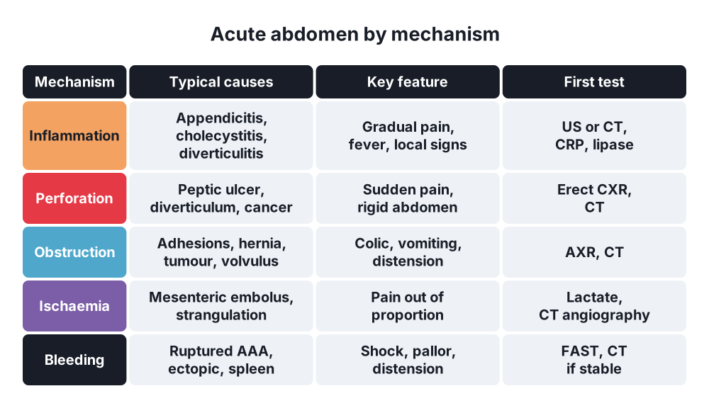
- Inflammation → appendicitis, cholecystitis, diverticulitis, pancreatitis, Meckel's diverticulitis, PID
- Perforation → peptic ulcer, diverticulum, colorectal cancer, Boerhaave's, ischaemic bowel, foreign body
- Obstruction → adhesions, hernias, tumour, volvulus, intussusception, gallstone ileus
- Ischaemia → mesenteric arterial or venous occlusion, NOMI, strangulated hernia, volvulus, ischaemic colitis, torsion (ovary, testis, omentum)
- Bleeding → ruptured AAA, ruptured ectopic pregnancy, ruptured spleen or liver tumour, haemorrhagic ovarian cyst
- Trauma → solid organ or hollow viscus injury
By region (where the pain is)
- Right upper quadrant → cholecystitis, biliary colic, cholangitis, hepatitis, liver abscess, perforated duodenal ulcer, right lower lobe pneumonia
- Epigastrium → peptic ulcer, pancreatitis, gastritis, myocardial infarction, AAA
- Left upper quadrant → splenic infarct or rupture, pancreatitis, left lower lobe pneumonia
- Right iliac fossa → appendicitis, mesenteric adenitis, Meckel's, Crohn's, caecal diverticulitis, ectopic pregnancy, ovarian torsion or cyst, ureteric colic
- Left iliac fossa → sigmoid diverticulitis, colitis, sigmoid volvulus, ectopic, ovarian pathology, ureteric colic
- Central or diffuse → early appendicitis, small bowel obstruction, mesenteric ischaemia, peritonitis, AAA, gastroenteritis, DKA
- Loin to groin → renal colic, pyelonephritis, AAA
Non-surgical causes
- Cardiopulmonary → myocardial infarction, pneumonia, pulmonary embolism, pericarditis
- Metabolic → diabetic ketoacidosis, hypercalcaemia, uraemia, Addison's crisis, acute porphyria
- Haematological → sickle cell crisis, Henoch–Schönlein purpura, leukaemia
- Infections → gastroenteritis, spontaneous bacterial peritonitis, herpes zoster
- Drugs and toxins → lead poisoning, opioid withdrawal, cocaine
- Other → familial Mediterranean fever, abdominal migraine, irritable bowel syndrome
4. Pathogenesis
- Visceral pain → autonomic fibres from organ walls; dull, crampy, poorly localised to the midline by embryology:
- Foregut (stomach to second part of duodenum, liver, gallbladder, pancreas) → epigastrium
- Midgut (to two-thirds of the transverse colon) → periumbilical
- Hindgut (to the upper anal canal) → suprapubic
- Parietal (somatic) pain → somatic nerves of the parietal peritoneum; sharp, well localised, worse with movement and coughing
- Referred pain → felt at a distance from the organ, along shared nerve roots:
- Diaphragm (C3–5) → shoulder tip (Kehr's sign with splenic rupture)
- Biliary → right scapula
- Pancreas → back
- Ureter → groin, testis or labium
- Shifting pain → appendicitis starts periumbilical (visceral) and moves to the right iliac fossa (parietal)
- Colicky pain → obstruction of a hollow tube (bowel, ureter, gallbladder neck)
- Constant pain → inflammation, ischaemia, peritonitis
- Pain out of proportion to the findings → ischaemia (acute mesenteric ischaemia)
- Peritonitis → fluid loss, ileus, bacterial translocation, sepsis, multi-organ failure
5. Classification
By main mechanism (the "5 Is" and more)
- Inflammatory, perforative, obstructive, ischaemic (vascular), haemorrhagic, traumatic
By urgency
- Immediate surgery (minutes) → unstable ruptured AAA, haemoperitoneum with shock, ruptured ectopic
- Urgent surgery (hours) → perforation with peritonitis, strangulated obstruction, mesenteric ischaemia, necrotising infection
- Expedited (within 24 hours) → appendicitis, uncomplicated obstruction failing conservative care, cholecystitis
- Conservative first → uncomplicated diverticulitis, pancreatitis, adhesive obstruction without strangulation, biliary colic
NCEPOD classification of intervention
- Immediate (minutes), urgent (hours), expedited (days), elective
6. Clinical picture
History
- Pain (SOCRATES) → site, onset, character, radiation, associated symptoms, timing, exacerbating and relieving factors, severity
- Sudden → perforation, rupture, torsion, mesenteric embolus
- Gradual → inflammation
- Vomiting → before pain in gastroenteritis; after pain in surgical disease; bilious or faeculent in obstruction
- Bowel habit → absolute constipation (obstruction), diarrhoea (gastroenteritis, ischaemic colitis)
- Urinary symptoms, menstrual history (last period, contraception, risk of pregnancy)
- Past history → previous surgery (adhesions), AF and vascular disease (ischaemia), gallstones, alcohol, NSAIDs or steroids (ulcer)
- Drugs → anticoagulants, steroids, immunosuppression (masks signs)
Examination
- General → pallor, sweating, jaundice, dehydration, posture (peritonitis lies still; colic writhes)
- Observations → pulse, BP, respiratory rate, temperature, SpO2, urine output, capillary refill; look for sepsis and shock
- Abdomen:
- Inspection → distension, scars, hernias, bruising (Cullen's, Grey Turner's)
- Palpation → tenderness, guarding, rigidity, rebound, masses, pulsatile mass (AAA)
- Percussion → tympany (obstruction), loss of liver dullness (free air), shifting dullness, percussion tenderness
- Auscultation → silent (ileus, peritonitis) or tinkling (obstruction); limited value
- Hernial orifices (always), external genitalia (torsion), DRE, pelvic examination when indicated
- Chest → pneumonia, effusion, heart signs
Eponymous signs
- Murphy's → stops breathing in when the gallbladder is palpated (cholecystitis)
- Rovsing's → pressure in the left iliac fossa causes pain on the right (appendicitis)
- Psoas and obturator signs → retrocaecal and pelvic appendix
- Blumberg's → rebound tenderness (peritonitis)
- Shchetkin–Blumberg (Bulgarian and Russian texts) → same as rebound tenderness
- Carnett's → pain gets worse when the abdominal wall is tensed (abdominal wall cause)
- Kehr's → left shoulder tip pain (splenic rupture)
- Cullen's and Grey Turner's → periumbilical and flank bruising (pancreatitis, retroperitoneal bleeding)
7. Diagnosis
Bedside
- Observations and early warning score (NEWS2); urinalysis; pregnancy test in every woman of childbearing age; capillary glucose; ECG
Blood tests
- FBC (white cells, haemoglobin), CRP, U&E, LFTs, amylase or lipase, calcium, glucose, clotting
- Blood gas and lactate → sepsis, ischaemia, acidosis
- Group and save or crossmatch; blood cultures if febrile
Imaging
- Erect chest X-ray → free air under the diaphragm (perforation; absent in about 20–30%)
- Abdominal X-ray → obstruction, volvulus (coffee bean sign), stones; low yield otherwise
- Ultrasound → gallbladder, biliary tree, free fluid, gynaecological and renal causes, AAA; FAST in trauma; first choice in pregnancy and children
- CT abdomen and pelvis with IV contrast → the most useful single test in adults; accuracy over 90%; guides surgery
- CT angiography → suspected mesenteric ischaemia, bleeding, AAA
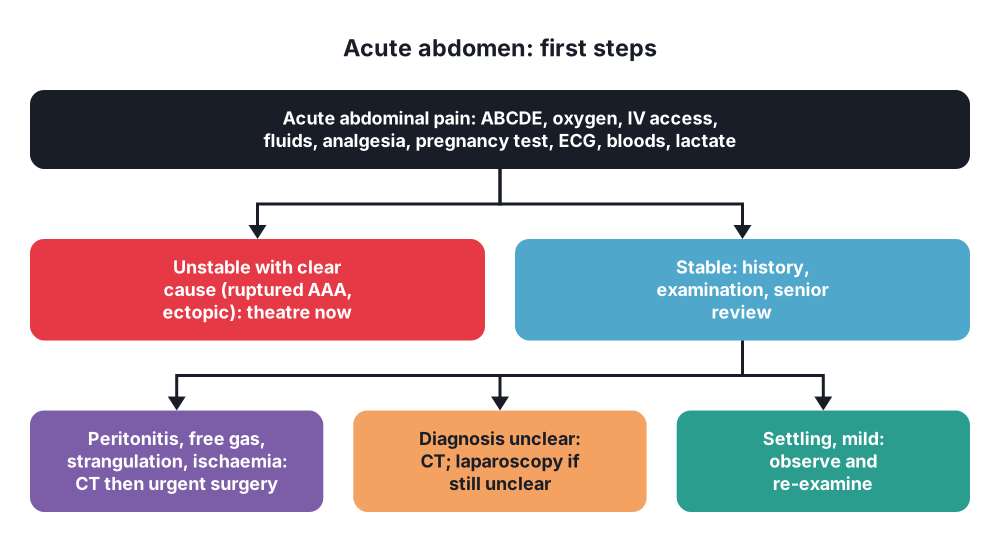
- Do not delay surgery for imaging in an unstable patient with a clear diagnosis (ruptured AAA with shock goes to theatre)
- MRI → pregnancy (appendicitis), biliary tree (MRCP)
Invasive
- Diagnostic laparoscopy → when the diagnosis is still unclear, especially young women with right iliac fossa pain; can be therapeutic
- Diagnostic peritoneal lavage → largely replaced by FAST and CT
Scores
- Alvarado and AIR score (appendicitis), Glasgow-Imrie (pancreatitis), qSOFA (sepsis)
- NELA risk calculator or P-POSSUM before emergency laparotomy
8. Differential diagnosis
- Surgical against medical causes (see section 3)
- In women → ectopic pregnancy, ovarian torsion, ruptured or haemorrhagic cyst, PID, endometriosis, mittelschmerz
- In the elderly → mesenteric ischaemia, AAA, perforated cancer, volvulus; fewer signs, less fever, less leucocytosis
- In children → intussusception, malrotation with volvulus, mesenteric adenitis, testicular torsion, Hirschsprung's enterocolitis
- In pregnancy → appendicitis (pain higher), cholecystitis, placental abruption, uterine rupture, HELLP syndrome
- Immunosuppressed → neutropenic enterocolitis, CMV colitis, fewer signs
- Postoperative → anastomotic leak, collection, missed injury
9. Treatment
Conservative
- Resuscitate first (ABCDE):
- High-flow oxygen, two large-bore cannulas
- IV fluids (balanced crystalloid), blood if bleeding
- Urinary catheter and fluid balance
- NG tube if vomiting or obstructed
- Analgesia early → IV paracetamol and titrated opioids; analgesia does not mask the diagnosis and improves examination
- Antiemetics
- Sepsis → antibiotics within 1 hour (sepsis bundle), cultures first if possible
- Nil by mouth if surgery is likely; VTE prophylaxis; stop or reverse anticoagulants as needed
- Senior review and early involvement of anaesthetics and critical care in high-risk patients
- Active observation with repeated examination → for doubtful cases (non-specific pain, possible appendicitis)
- Conservative management of selected conditions:
- Uncomplicated diverticulitis
- Pancreatitis
- Adhesive small bowel obstruction (water-soluble contrast)
- Uncomplicated appendicitis in selected patients (antibiotics)
- Interventional radiology → percutaneous drainage of abscesses, embolisation of bleeding, endovascular repair of AAA, mesenteric stenting
Surgical
- Indications for urgent operation:
- Generalised peritonitis
- Free gas with clinical deterioration
- Strangulated or closed-loop obstruction, failure of conservative treatment
- Ischaemic bowel, mesenteric ischaemia
- Uncontrolled bleeding (haemoperitoneum, ruptured ectopic, ruptured AAA)
- Clinical deterioration despite resuscitation
- Laparoscopy → diagnostic and therapeutic in stable patients (appendicitis, cholecystitis, perforated ulcer)
- Laparotomy → midline; full exploration; deal with the cause; peritoneal lavage
- Damage control surgery → for the physiologically exhausted patient (acidosis, hypothermia, coagulopathy): control bleeding and contamination, temporary closure, ICU, return in 24–48 hours
- Perioperative care pathway (NELA standards) → consultant surgeon and anaesthetist present for high-risk cases, theatre within the time set by urgency, ICU after surgery if predicted mortality over 5%, review of older patients by a geriatrician
10. Complications
- Sepsis and septic shock, multi-organ failure
- Hypovolaemic shock from bleeding or third-space losses
- Missed diagnosis → perforation, gangrene, death (especially elderly, pregnant, immunosuppressed)
- Negative laparotomy or laparoscopy (normal findings)
- Surgical complications → wound infection, anastomotic leak, intra-abdominal collection, ileus, abdominal compartment syndrome, VTE, pneumonia
Prognosis
- Depends on cause, age, frailty and delay; NELA 30-day mortality for emergency laparotomy about 9–10%, over 20% in the over-80s
Show as a list
Acute abdomen
- Mechanisms
- Inflammation
- Perforation
- Obstruction
- Ischaemia
- Bleeding
- Pain
- Visceral: midline
- Parietal: localised
- Referred
- Out of proportion: ischaemia
- Assessment
- History, exam, hernias, DRE
- Pregnancy test, ECG
- Bloods, lactate
- Imaging
- Erect CXR
- Ultrasound
- CT with contrast
- Management
- ABCDE, fluids
- Early analgesia
- Antibiotics in 1 h
- Theatre if peritonitis
- Laparoscopy if unclear
- Mimics
- MI, DKA, pneumonia


