
87. Abdominal wall and abdominal organ trauma
High yield
Blunt and penetrating injury to the abdominal wall, hollow organs and retroperitoneum; FAST, CT, laparotomy and damage control.
Exam Prep · State examSurgery · General surgerySurgery · Emergency and trauma
1. Definition
- Abdominal trauma → injury to the abdominal wall, the peritoneal cavity and its organs, or the retroperitoneum
- Closed (blunt) → the skin is intact; force passes through the wall (road traffic collision, fall, blow, crush)
- Open (penetrating) → a wound passes through the skin; stab wounds, gunshot wounds, impalement
- Non-penetrating wound → stops in the wall; the peritoneum is intact
- Penetrating wound → breaches the parietal peritoneum; may or may not injure organs
- Isolated wall injury → contusion, haematoma, muscle tear, traumatic hernia without organ injury
- Organ injury may be to solid organs (liver, spleen, kidney, pancreas: bleed) or hollow organs (stomach, duodenum, small bowel, colon, rectum, bladder: leak and peritonitis)
- Liver and biliary trauma → question 69; pancreas → question 74; spleen → question 79; multiple injuries → question 88
2. Epidemiology
- Abdominal injury in about 10–15% of major trauma; a common cause of preventable trauma death (missed bleeding)
- Blunt trauma → most common in Europe (about 80%); road traffic collisions cause most
- Penetrating trauma → stab wounds more common than gunshot wounds in Europe; gunshot wounds dominate in the USA and South Africa
- Most injured organs:
- Blunt → spleen, then liver, then kidney; hollow organs in only about 1% (small bowel and its mesentery most often)
- Stab wounds → liver, small bowel, diaphragm, colon
- Gunshot wounds → small bowel, colon, liver, abdominal vessels
- Hollow organ injury is the most often missed abdominal injury
- Duodenal injuries → under 2% of abdominal trauma; about 80% from gunshot wounds; 70% have other abdominal injuries
- Colon injury mortality → 22–35% in World War II (mandatory colostomy) vs about 1% today
3. Aetiology and risk factors
Mechanisms of blunt injury
- Direct blow (compression) → organ crushed between the wall and the spine (pancreas, duodenum, small bowel, kidney)
- Sudden rise in pressure (burst) → a closed loop of bowel or the stomach bursts, usually on the antimesenteric border
- Deceleration (shear) → fixed and mobile parts move at different speeds → mesenteric tears, renal pedicle, liver at its ligaments, duodenojejunal flexure
- Seat belt (lap belt) → bowel and mesentery crushed against the spine; associated Chance fracture of the lumbar spine (L1–L3)
- Handlebar (children on bicycles) → duodenum, pancreas
Penetrating injury
- Stab wounds → low energy; injure only what the blade touches; about two thirds penetrate the peritoneum, and only about half of these injure an organ
- Gunshot wounds → high energy; unpredictable track; cavitation; about 80–90% injure an organ
- Shotgun, blast and fragment injuries → many wounds
Risk markers for abdominal injury
- High-energy mechanism, ejection, death in the same vehicle, pedestrian struck
- Seat belt sign, fractures of the lower ribs (liver, spleen), pelvic fracture, lumbar spine fracture (Chance)
- Altered consciousness (head injury, alcohol, drugs), spinal cord injury, distracting injuries → examination unreliable
4. Pathogenesis
Two ways an abdominal injury kills
- Haemorrhage (solid organs, mesentery, vessels) → hypovolaemic shock; early death
- Contamination (hollow organs) → chemical then bacterial peritonitis, sepsis; later death
Hollow organ injury
- Stomach → acid causes rapid, strong peritoneal signs; must open the lesser sac to see the posterior wall
- Duodenum → mostly retroperitoneal; leak into the retroperitoneum gives few early signs; intramural haematoma may cause gastric outlet obstruction (children)
- Small bowel → perforation, serosal tears, bucket-handle mesenteric tears (devascularised bowel), late stricture from ischaemia
- Colon → contamination with faeces; retroperitoneal parts (ascending, descending) may leak without peritonitis
- Rectum → intraperitoneal (treated like colon) or extraperitoneal (pelvic sepsis); penetrating wounds, pelvic fracture fragments, impalement
Abdominal wall
- Contusion and haematoma → bruising, pain; may hide deeper injury
- Rectus sheath haematoma → torn inferior epigastric vessels (trauma, coughing, anticoagulants); below the arcuate line the haematoma can spread into the pelvis
- Traumatic abdominal wall hernia → muscles and fascia torn but the skin intact (handlebar, seat belt); bowel may herniate under the skin
- Degloving (Morel-Lavallée lesion) → skin and fat sheared off the fascia; fluid collection
Retroperitoneal haematoma
- From the kidney, pancreas, duodenum, major vessels, or pelvic fracture (most common blunt cause)
- Can hold several litres of blood with few signs
5. Classification
By mechanism
- Blunt (closed) and penetrating (open) → stab, gunshot (low or high velocity), shotgun, blast, impalement
By injured structure
- Abdominal wall only
- Solid organ (bleeding), hollow organ (perforation), vascular (major vessels, mesentery), combined
AAST organ injury scales (grades I–V or VI)
- Small bowel:
- I → contusion or haematoma, or partial-thickness laceration
- II → laceration under 50% of the circumference
- III → laceration 50% or more of the circumference without transection
- IV → transection
- V → transection with segmental tissue loss, or devascularised segment
- Colon → similar grades I–V; Sabiston groups them into non-destructive (under 50% of the circumference) and destructive (50% or more, transection, devascularised)
- Rectum → destructive if over 25% of the circumference
- Duodenum → I haematoma of one part or partial-thickness tear; II larger haematoma or tear under 50%; III tear 50–100% of D2 or 50–75% of D1, D3, D4; IV disruption over 75% of D2 or the ampulla or distal bile duct; V massive disruption of the duodenopancreatic complex or devascularisation
Retroperitoneal zones
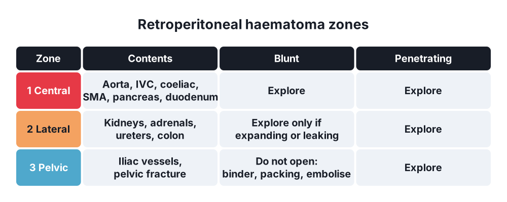
- Zone 1 (central) → aorta, IVC, coeliac, superior mesenteric vessels, pancreas, duodenum
- Zone 2 (lateral, flanks) → kidneys, adrenals, ureters, colon
- Zone 3 (pelvic) → iliac vessels, pelvic fracture bleeding
6. Clinical picture
History
- Mechanism → speed, seat belt, airbag, ejection, fall height; weapon type, number of shots, direction
- Time of injury, last meal, drugs (anticoagulants), alcohol
Symptoms
- Abdominal pain (may be mild early, or masked by other injuries, head injury or alcohol)
- Shoulder tip pain → blood under the diaphragm (Kehr's sign on the left in splenic injury)
- Nausea, vomiting, haematemesis (stomach or duodenum), haematuria (kidney, bladder)
Signs
- Shock → tachycardia, hypotension, cold periphery, low urine output (bleeding)
- Peritonitis → tenderness, guarding, rigidity, rebound, absent bowel sounds (hollow organ leak)
- Fresh blood is only a mild irritant; a soft abdomen does not exclude major bleeding
- Distension → late sign of major bleeding
- Seat belt sign → bruising across the lower abdomen; several times higher risk of bowel, mesenteric and lumbar spine (Chance) injury
- Cullen's (periumbilical) or Grey Turner's (flank) bruising → retroperitoneal bleeding
- Wounds → inspect the front, flanks, back, perineum and buttocks (log-roll); mark each wound
- Evisceration of omentum or bowel through a wound
- Rectal and vaginal examination → blood (rectal or vaginal injury), high-riding prostate (urethral injury), bone fragments
- Rectus sheath haematoma → tender mass that does not cross the midline and stays palpable when the patient tenses the abdomen (Fothergill's sign)
Duodenal and pancreatic injury
- Few signs early; back pain, rising amylase, delayed peritonitis or sepsis after 24–48 hours
7. Diagnosis
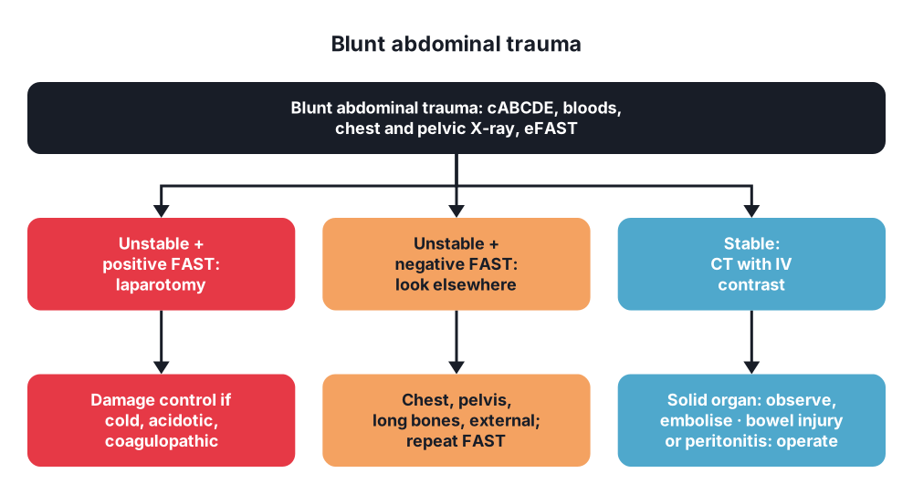
- Start with the ATLS primary survey (see question 88); the aim is to find bleeding that needs surgery
- Clinical examination alone is unreliable in trauma (head injury, intoxication, distracting injuries)
Bloods
- FBC (first haemoglobin may be normal), group and crossmatch, coagulation, blood gas: lactate and base deficit, U&E, amylase (late and non-specific), LFT
- Urinalysis → haematuria (kidney or bladder); pregnancy test
Imaging and bedside tests
- eFAST (extended focused assessment with sonography for trauma):
- Four views → pericardium, hepatorenal (Morison's pouch), splenorenal, pelvis (rectovesical or rectouterine pouch); extended → both pleural spaces (pneumothorax, haemothorax)
- Shows free fluid (about 100–250 mL or more); quick, bedside, repeatable
- Unstable + positive FAST → laparotomy
- Poor for hollow organ, retroperitoneal, pancreatic and diaphragmatic injury
- CT with IV contrast → gold standard in stable patients:
- Grades solid organ injury, shows active contrast extravasation (blush), retroperitoneum, spine and pelvis
- Hollow organ injury → free fluid without solid organ injury, bowel wall thickening, mesenteric stranding or haematoma, free gas, abrupt loss of wall enhancement; sensitivity only 55–95%
- Unexplained free fluid → repeat examination, admission, often laparoscopy or laparotomy
- Triple contrast (oral, rectal, IV) for back and flank stab wounds in some centres
- Diagnostic peritoneal lavage (DPL) or aspiration → largely replaced by FAST; used where FAST is not available or is equivocal in an unstable patient:
- Positive → 10 mL of free blood on aspiration, red cells over 100,000/mm³, white cells over 500/mm³, bile, bacteria, food fibres or amylase in the fluid
- Misses retroperitoneal injury; too sensitive (leads to non-therapeutic laparotomy)
- Chest X-ray (free gas under the diaphragm, haemothorax, diaphragmatic injury) and pelvic X-ray (pelvic fracture) in the resuscitation room
- Diagnostic laparoscopy → stable patients with stab wounds to the left thoracoabdominal area (diaphragmatic injury) or doubtful peritoneal penetration
- Local wound exploration → anterior stab wounds in stable patients without peritonitis; if the posterior fascia is intact, the patient may go home
- Water-soluble contrast study (upper GI series) or CT with oral contrast → suspected duodenal injury
- Retrograde urethrogram before a catheter if there is blood at the meatus; CT cystogram for bladder injury
- Rigid sigmoidoscopy under anaesthesia → suspected rectal injury
- Serial clinical examination by the same surgeon, ideally every 4–6 hours, in patients under observation
Indications for immediate laparotomy
- Haemodynamic instability with a positive FAST or DPL
- Peritonitis
- Gunshot wound with peritoneal penetration (most centres; selective in experienced centres)
- Evisceration of bowel, impaled object (removed in theatre)
- Free gas on imaging, diaphragmatic rupture
- Blood from the stomach (NG tube) or the rectum after a penetrating wound
8. Differential diagnosis
- Extra-abdominal bleeding causing shock → chest (haemothorax), pelvic fracture, long bone fractures, external bleeding
- Other causes of shock → tension pneumothorax, cardiac tamponade, spinal shock (bradycardia, warm), cardiac contusion
- Referred abdominal pain → lower rib fractures, pulmonary contusion, spinal fracture
- Abdominal wall haematoma vs intraperitoneal injury
- Retroperitoneal haematoma (pelvic fracture) vs intraperitoneal bleeding → a false-positive DPL
- Pre-existing disease → ruptured aneurysm, perforated ulcer (the "fall" may follow collapse)
9. Treatment
Conservative
- Resuscitation (see question 88) → cABCDE, two large-bore cannulas, massive transfusion protocol (red cells, plasma, platelets 1:1:1), tranexamic acid within 3 hours, permissive hypotension (systolic about 80–90 mmHg until bleeding is controlled; not with head injury), warm fluids, avoid large volumes of crystalloid
- Selective non-operative management (SNOM):
- Blunt solid organ injury in stable patients → observation, serial examination and haemoglobin, angioembolisation for contrast blush (see questions 69 and 79)
- Stab wounds in stable patients without peritonitis → serial examination for 24 hours; about 50% avoid surgery
- Selected gunshot wounds (tangential, right upper quadrant liver injury) in stable patients → CT and observation in experienced centres
- Needs a surgeon, CT, theatre and ICU available at all times
- Stop SNOM → instability, peritonitis, falling haemoglobin despite transfusion
- Duodenal haematoma → NG decompression, parenteral nutrition, contrast study at 5–7 days; surgery if the obstruction lasts over about 14 days
- Rectus sheath haematoma → rest, analgesia, reverse anticoagulation; embolisation of the inferior epigastric artery if it keeps bleeding
- Abdominal wall contusion → analgesia; look for underlying injury
- Antibiotics → single preoperative dose for penetrating injury; no more than 24 hours after surgery, even with hollow organ injury
- Tetanus prophylaxis for wounds
Surgical
- Trauma laparotomy:
- Supine, arms out, prepped from the chin to the knees (allows thoracotomy and groin access)
- Long midline incision (xiphoid to pubis)
- Steps:
- Remove blood and clot; pack all four quadrants
- Control bleeding first (packing, pressure, clamps, Pringle manoeuvre); aortic compression at the hiatus if needed
- Control contamination (clamps, staples, quick sutures)
- Systematic inspection → liver, spleen, stomach (front and back through the lesser sac), duodenum (Kocher manoeuvre), pancreas, whole small bowel from the duodenojejunal flexure to the ileocaecal valve (both sides of each loop and the mesentery), colon, rectum, bladder, diaphragm, retroperitoneum
- Definitive repair or damage control
- Stomach → debride and close in one or two layers (stapler possible)
- Duodenum:
- Most (about 98%) can be repaired primarily after debridement; wide Kocher manoeuvre; omental patch
- Large tissue loss away from the ampulla → resection and duodenoduodenostomy or Roux-en-Y duodenojejunostomy
- Complex injuries → pyloric exclusion with gastrojejunostomy, duodenal diverticulisation or tube duodenostomy (now used selectively); pancreaticoduodenectomy only for destroyed duodenopancreatic complex, usually staged after damage control
- Intramural haematoma found at surgery → do not open unless full-thickness injury is suspected
- Small bowel:
- Serosal tear → interrupted seromuscular sutures
- Perforation under 50% of the circumference → debride and close transversely (avoids narrowing)
- Over 50% of the circumference, several close perforations, or devascularised segment → resection and anastomosis (stapled and hand-sewn equal)
- Mesenteric tear → tear parallel to the bowel (bucket handle) usually devascularises the bowel → resect; tear at right angles to the bowel → suture the mesentery; any bowel with poor perfusion → resect
- Colon:
- Non-destructive injury → primary repair in all patients (no colostomy)
- Destructive injury → resection with primary anastomosis in stable patients without major comorbidity; consider a stoma with shock, over 6 units of blood, major comorbidity, immunosuppression or an open abdomen
- Damage control → resect, staple off the ends, delayed anastomosis at the second look
- Sabiston: leak rate 42% in transfused, comorbid patients after resection vs 3% in healthy patients
- Rectum:
- Intraperitoneal → manage as colon (repair or resection)
- Extraperitoneal → faecal diversion (loop colostomy or ileostomy) for destructive injuries; routine presacral drainage and distal washout no longer recommended; contrast enema before stoma closure at about 6 weeks
- Abdominal wall:
- Traumatic abdominal wall hernia → repair, often at laparotomy (look for bowel injury); delayed repair with mesh if contaminated
- Debride devitalised muscle and fascia; close the fascia
- Morel-Lavallée lesion → aspiration, compression, or debridement and drainage
- Retroperitoneal haematoma at laparotomy:
- Zone 1 → always explore (blunt or penetrating); medial visceral rotation (Mattox on the left, Cattell-Braasch on the right)
- Zone 2 → explore penetrating injuries; in blunt injury explore only if expanding, pulsatile or with urine leak
- Zone 3 → explore penetrating injuries; in blunt injury do not open (pelvic fracture bleeding: pelvic binder, preperitoneal packing, angioembolisation)
- Damage control surgery → for the "lethal diamond" (hypothermia, acidosis, coagulopathy, hypocalcaemia):
- Short operation (under 60–90 minutes): stop bleeding, control contamination, packing, temporary abdominal closure (vacuum dressing)
- ICU: rewarm, correct acidosis and coagulopathy
- Return to theatre in 24–48 hours: remove packs, definitive repair, anastomosis, close the abdomen
- Resuscitative thoracotomy or REBOA (balloon in the aorta) → selected patients in extremis
- Laparoscopy → diagnostic and therapeutic in stable patients (diaphragm repair, small bowel repair) in experienced hands
10. Complications
Of the injury
- Haemorrhagic shock, death
- Peritonitis, intra-abdominal abscess, sepsis, multiple organ failure
- Missed injuries (duodenum, pancreas, small bowel, diaphragm) → delayed peritonitis, mortality rises with each hour of delay
- Duodenal fistula, pancreatic fistula
- Abdominal compartment syndrome (see question 98)
- Late bowel stricture (ischaemic) → obstruction weeks later
- Traumatic diaphragmatic hernia, delayed splenic rupture
Of surgery
- Anastomotic or repair leak, enterocutaneous fistula
- Wound infection, wound dehiscence, incisional hernia
- Open abdomen → enteroatmospheric fistula, giant ventral hernia
- Adhesive bowel obstruction
- Stoma complications (if formed)
- Non-therapeutic laparotomy → complications in up to 40%
Prognosis
- Depends on shock on arrival, number of injured organs, associated injuries and time to haemorrhage control
Show as a list
Abdominal trauma
- Mechanism
- Blunt: crush, burst, shear
- Seat belt: bowel + Chance
- Stab: low energy
- Gunshot: high energy
- Organs
- Solid: bleed
- Hollow: leak
- Retroperitoneum: zones 1-3
- Wall: haematoma, hernia
- Tests
- eFAST: unstable
- CT: stable
- DPL: backup
- Laparoscopy: diaphragm
- Treatment
- cABCDE, MTP, TXA
- SNOM if stable
- Trauma laparotomy
- Damage control
- Hollow organs
- Bowel <50%: repair
- Bowel >50%: resect
- Colon: repair or resect
- Rectum: divert


