52. Anterior abdominal wall, hernias in general and congenital abdominal wall defects
High yield
Layers of the abdominal wall; parts, types and complications of a hernia; exomphalos and gastroschisis in the newborn.
Exam Prep · State examSurgery · General surgerySurgical Anatomy · Abdominal wall and hernia
1. Definition
- Hernia → abnormal protrusion of a viscus, or part of it, through a defect in the wall of the cavity that contains it
- External abdominal hernia → through the abdominal wall (groin, umbilicus, incision)
- Internal hernia → into a pocket or opening inside the abdomen
- Congenital abdominal wall defect → failure of the abdominal wall to close in the fetus: exomphalos (omphalocele) and gastroschisis
2. Epidemiology
- Hernias → about 5% of people; lifetime risk of a groin hernia 27–43% in men, 3–6% in women
- Frequency → inguinal (about 75%), then incisional, umbilical, femoral
- Groin hernias → third commonest cause of bowel obstruction in the West (after adhesions and cancer); commonest in many low-income countries
- Exomphalos → about 1 in 5,000–7,000 births
- Gastroschisis → about 1 in 4,000–7,000 births, rising; young mothers, smoking, drugs
3. Aetiology and risk factors
Anatomy of the anterior abdominal wall (revision)
- Layers from outside in:
- Skin
- Subcutaneous fat → Camper's (fatty) and Scarpa's (membranous) fascia below the umbilicus
- External oblique → fibres "hands in pockets"; aponeurosis forms the inguinal ligament
- Internal oblique
- Transversus abdominis
- Transversalis fascia
- Extraperitoneal fat
- Parietal peritoneum
- Rectus abdominis in the rectus sheath; linea alba in the midline; linea semilunaris at the lateral edge of rectus
- Arcuate line (about one-third of the way from umbilicus to pubis) → below it all aponeuroses pass in front of rectus; posterior sheath absent
- Blood supply → superior and inferior epigastric arteries (inside the sheath, behind rectus)
- Nerves → T7–T12, iliohypogastric and ilioinguinal (L1); run between internal oblique and transversus
- On the inside → median umbilical fold (urachus), medial folds (obliterated umbilical arteries), lateral folds (inferior epigastric vessels)
Causes of hernia
- Congenital → patent processus vaginalis (indirect inguinal), umbilical ring not closed
- Acquired → weak wall plus raised pressure:
- Raised pressure → chronic cough, constipation, straining to pass urine (prostate), heavy lifting, obesity, pregnancy, ascites, peritoneal dialysis
- Weak wall → age, smoking, collagen disorders (Ehlers-Danlos, Marfan's), previous incision, wound infection, nerve injury
Congenital wall defects
- Exomphalos → gut fails to return from the umbilical cord at week 10–12
- Gastroschisis → defect of the wall beside the cord, probably from a vascular accident
4. Pathogenesis
Parts of a hernia
- Defect (orifice) in the wall
- Sac → peritoneum: neck, body, fundus
- Contents → omentum (omentocele), small bowel (enterocele), others
- Coverings → layers of the wall pushed in front of the sac
Strangulation sequence
- Tight neck compresses the contents
- Venous and lymphatic return blocked → oedema
- Pressure rises → arterial inflow stops
- Ischaemia → gangrene → perforation → peritonitis, sepsis
- Bowel can die within 6 hours
5. Classification
By site
- Groin → inguinal (indirect, direct), femoral
- Ventral → umbilical, paraumbilical, epigastric, Spigelian, incisional
- Lumbar (Petit's inferior triangle, Grynfeltt's superior triangle), obturator, perineal, sciatic
- Internal → paraduodenal, foramen of Winslow, diaphragmatic
By state
- Reducible → contents go back
- Irreducible (incarcerated) → cannot be reduced, adhesions or narrow neck; blood supply intact
- Obstructed → bowel lumen blocked; blood supply intact
- Strangulated → blood supply cut off → urgent surgery
Special hernias
- Richter's → only part of the bowel wall is trapped; can strangulate without obstruction
- Littré's → contains a Meckel's diverticulum
- Amyand's → appendix in an inguinal hernia
- De Garengeot's → appendix in a femoral hernia
- Sliding (hernia en glissade) → a retroperitoneal organ (colon, bladder) forms part of the sac wall
- Maydl's (W-hernia) → two loops in the sac; the middle loop inside the abdomen strangulates
- Pantaloon → direct and indirect together, either side of the inferior epigastric vessels
Congenital defects
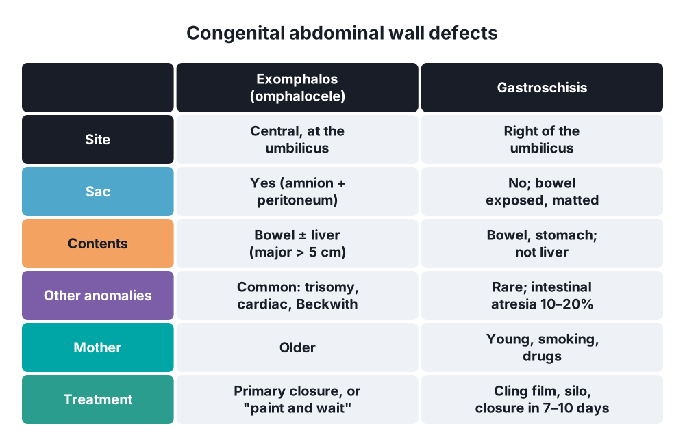
- Exomphalos minor (< 5 cm, bowel only) and major (> 5 cm, liver too)
6. Clinical picture
Symptoms
- Lump that appears on standing or straining and goes away on lying down
- Dragging ache, worse at the end of the day
- Strangulation → sudden severe pain, tender, tense, irreducible lump, no cough impulse; colicky pain, vomiting, distension, no flatus
Signs
- Examine standing and lying, both sides
- Cough impulse, reducibility
- Strangulation → red, hot, tender lump; tachycardia, fever; signs of obstruction or peritonitis
- Look for causes → chest, abdomen (masses, ascites), prostate, scars
Newborn defects
- Exomphalos → central defect at the umbilicus; contents in a membrane sac (amnion and peritoneum); cord inserts into the sac
- Gastroschisis → defect right of the umbilicus; no sac; bowel thick, oedematous and matted from exposure to amniotic fluid; cord normal
7. Diagnosis
- Hernia → clinical in most
- Ultrasound → small or uncertain groin hernia, occult hernia
- CT → obese patients, incisional hernia, obturator hernia, strangulation
- MRI → athletes' groin pain
- Blood tests in strangulation → FBC, U&E, lactate, group and save
- Abdominal X-ray → bowel obstruction
Congenital defects
- Antenatal ultrasound and raised maternal serum AFP
- Exomphalos → karyotype, echocardiography (cardiac defects), renal ultrasound, blood glucose (Beckwith-Wiedemann)
- Gastroschisis → look for intestinal atresia (10–20%); other anomalies rare
8. Differential diagnosis
- Groin lump → see inguinal and femoral hernia: lymph node, saphena varix, femoral aneurysm, lipoma, hydrocele, undescended testis, psoas abscess
- Ventral lump → lipoma, divarication of the recti (no defect, no risk of strangulation), desmoid, rectus sheath haematoma
- Strangulated hernia versus torsion of the testis, inguinal lymphadenitis
9. Treatment
Conservative
- Watchful waiting → small, minimally symptomatic inguinal hernia in men, or patients unfit for surgery
- Correct risk factors → stop smoking, weight loss, treat cough and constipation, prostate
- Truss → little value; not for femoral hernias
- Never try forceful reduction (taxis) of a strangulated hernia → "reduction en masse" hides dead bowel
Surgical: principles of hernia repair
- Herniotomy → dissect, open, inspect, reduce and remove the sac (children: herniotomy alone)
- Herniorrhaphy → sutured repair of the defect (Shouldice, McVay, Mayo)
- Hernioplasty → tension-free mesh reinforcement (Lichtenstein, TEP, TAPP) → standard in adults
- Mesh → polypropylene (permanent); biological or absorbable mesh in contaminated fields
- Mesh positions → onlay, inlay, sublay (retromuscular or preperitoneal), intraperitoneal (IPOM)
- Open or laparoscopic; local, regional or general anaesthesia
Surgical: strangulated hernia
- Resuscitate → IV fluids, NG tube, catheter, analgesia, antibiotics
- Emergency operation
- Open the sac first, hold the contents (to avoid them slipping back unseen)
- Release the constriction
- Check viability → colour, peristalsis, mesenteric pulsation; wrap in warm saline-soaked swabs for 5–10 min
- Non-viable bowel → resection and anastomosis
- Repair → mesh if clean; sutured or biological mesh if contaminated
Surgical: congenital defects
- Gastroschisis:
- Cover the bowel in cling film, NG tube, IV fluids (large losses), warmth, antibiotics
- Primary reduction and closure, or preformed silo with gradual reduction over 7–10 days, then closure
- Parenteral nutrition until the gut works (weeks)
- Exomphalos:
- Protect the sac; look for other anomalies
- Minor → primary closure
- Major → paint the sac (silver sulfadiazine) to let it epithelialise ("paint and wait"); repair the ventral hernia later (about 1 year)
10. Complications
- Hernia → incarceration, obstruction, strangulation, perforation, peritonitis
- Repair → haematoma, seroma, wound infection, recurrence, chronic pain, mesh infection, injury to bowel, bladder or vessels
- Gastroschisis → short bowel, long parenteral feeding, sepsis, necrotising enterocolitis; survival > 90%
- Exomphalos → prognosis depends on associated anomalies (chromosomal, cardiac)
Show as a list
Hernias in general
- Abdominal wall
- Three flat muscles
- Rectus sheath
- Arcuate line
- Transversalis fascia
- Hernia parts
- Defect
- Sac
- Contents
- Coverings
- States
- Reducible
- Incarcerated
- Obstructed
- Strangulated
- Special types
- Richter's
- Littré's
- Amyand's
- Sliding
- Repair
- Herniotomy
- Herniorrhaphy
- Mesh hernioplasty
- Congenital
- Exomphalos: sac
- Gastroschisis: no sac
- Silo, closure


