
98. Abdominal compartment syndrome
High yield
Sustained intra-abdominal pressure over 20 mmHg with new organ failure; measure bladder pressure, treat medically, then decompress.
Exam Prep · State examSurgery · General surgerySurgery · Emergency and trauma
1. Definition
- Definitions of the World Society of the Abdominal Compartment Syndrome (WSACS, 2013):
- Intra-abdominal pressure (IAP) → steady-state pressure in the abdominal cavity; normal 5–7 mmHg in critically ill adults (about 0 in healthy people; higher in obesity and pregnancy)
- Intra-abdominal hypertension (IAH) → sustained or repeated IAP of 12 mmHg or more
- Abdominal compartment syndrome (ACS) → sustained IAP over 20 mmHg (with or without APP under 60 mmHg) with new organ dysfunction or failure
- Abdominal perfusion pressure (APP) → MAP minus IAP; target 60 mmHg or more
- Primary ACS → caused by injury or disease in the abdomen and pelvis (trauma, ruptured AAA, pancreatitis, peritonitis)
- Secondary ACS → from conditions outside the abdomen (massive fluid resuscitation, burns, sepsis, capillary leak)
- Recurrent ACS → returns after earlier treatment (including in an open abdomen)
2. Epidemiology
- IAH in about 30–50% of ICU patients; ACS in about 2–10%
- Common in → damage control trauma, ruptured AAA repair (about 10–20%), severe acute pancreatitis, major burns (over 30% TBSA), septic shock with large fluid volumes, liver transplant
- Mortality of established ACS → about 40–60% (up to 100% if not decompressed)
- Less common now in trauma because of damage control resuscitation (less crystalloid) and temporary abdominal closure
3. Aetiology and risk factors
Reduced abdominal wall compliance
- Abdominal surgery with tight fascial closure, major trauma, burns with eschar on the trunk
- Prone positioning, obesity, high head of bed (over 30°)
Increased intraluminal contents
- Ileus, gastroparesis, bowel obstruction, volvulus, pseudo-obstruction (Ogilvie's), toxic megacolon
Increased intra-abdominal contents
- Haemoperitoneum, retroperitoneal haematoma (pelvic fracture, ruptured AAA)
- Ascites (cirrhosis, large-volume fluids), peritoneal dialysis
- Intra-abdominal infection, abscess
- Tumours, packs left after damage control surgery, pneumoperitoneum (laparoscopy)
- Acute pancreatitis (oedema, fluid collections)
Capillary leak and fluid resuscitation
- Positive fluid balance (over 5 L in 24 hours), massive transfusion (over 10 units of blood in 24 hours)
- Sepsis, burns, pancreatitis, acidosis (pH under 7.2), hypothermia, coagulopathy
Other
- Mechanical ventilation with high PEEP, liver dysfunction, oliguria, hypotension
4. Pathogenesis
- The abdomen is a closed box with limited compliance; once the wall is stretched, small increases in volume cause steep rises in pressure
- Cardiovascular:
- IVC compression → less venous return → less preload → low cardiac output
- Raised systemic vascular resistance; diaphragm pushed up raises intrathoracic pressure
- Falsely high CVP and wedge pressure (misleading as a fluid guide)
- Respiratory:
- Diaphragm elevated → lower lung compliance, atelectasis
- High peak and plateau airway pressures, hypoxia, hypercapnia, hard to ventilate
- Renal:
- Renal vein compression and reduced renal perfusion → oliguria then anuria, acute kidney injury (one of the earliest signs; at about 15–20 mmHg)
- Renal perfusion pressure ≈ MAP − 2 × IAP
- Gut:
- Reduced mesenteric blood flow → gut ischaemia, mucosal acidosis, bacterial translocation, more oedema → a vicious cycle (oedema raises IAP)
- Liver → reduced hepatic blood flow, raised lactate (poor clearance)
- Brain:
- Raised intrathoracic pressure impairs venous outflow from the head → raised intracranial pressure (important with head injury)
- Abdominal wall → reduced blood flow, wound problems, ischaemia
- The final result is multiple organ dysfunction; untreated ACS is fatal
5. Classification
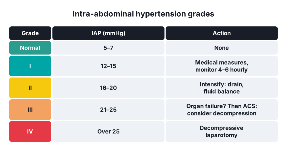
IAH grades (WSACS)
- Grade I → IAP 12–15 mmHg
- Grade II → IAP 16–20 mmHg
- Grade III → IAP 21–25 mmHg
- Grade IV → IAP over 25 mmHg
ACS by cause
- Primary (abdominal or pelvic cause; often needs early surgery or radiology)
- Secondary (outside the abdomen; fluid, sepsis, burns)
- Recurrent
Open abdomen (Björck classification)
- 1 → clean (1a not fixed, 1b fixed), 2 → contaminated, 3 → enteric leak, 4 → frozen abdomen with fistula
6. Clinical picture
- Usually a ventilated ICU patient after trauma, major surgery, pancreatitis, burns or massive resuscitation
- Clinical examination is unreliable for estimating IAP (sensitivity about 40–60%) → measure it
Signs
- Tense, distended abdomen
- Oliguria or anuria despite fluids
- Rising peak airway pressures, falling tidal volumes, hypoxia and hypercapnia
- Hypotension, tachycardia, rising vasopressor needs, raised CVP (misleading)
- Rising lactate, metabolic acidosis
- Raised ICP (head injury)
- Leg oedema, poor peripheral perfusion
7. Diagnosis
Measuring IAP
- Intravesical (bladder) pressure → the standard method:
- Patient supine, at end-expiration, abdominal muscles relaxed (sedation)
- Transducer zeroed at the mid-axillary line at the level of the iliac crest
- Instil up to 25 mL of sterile saline into the bladder through the catheter, clamp, wait 30–60 seconds, measure (in mmHg, not cmH2O)
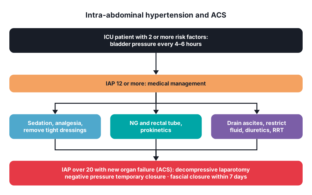
- Repeat every 4–6 hours in at-risk patients (or continuously)
- Other methods → intragastric, direct intraperitoneal (catheter), inferior vena cava
- Screen every ICU patient with two or more risk factors for IAH (WSACS)
Other tests
- APP (MAP − IAP), urine output, ventilator pressures, lactate, U&E (creatinine), blood gas
- CT (if the patient can travel) → round belly sign (AP to transverse diameter ratio over 0.8), compressed IVC, renal compression, bowel wall thickening, the cause (haematoma, collection, ascites)
- Ultrasound → ascites or collections that can be drained
8. Differential diagnosis
- Other causes of oliguria → hypovolaemia, sepsis-related AKI, nephrotoxins, urinary obstruction (blocked catheter)
- Other causes of high airway pressure → tension pneumothorax, bronchospasm, ARDS, tube problems
- Ongoing bleeding (haemoperitoneum) or a missed source of sepsis
- Mesenteric ischaemia (can cause and result from ACS)
- Cardiac failure, tamponade
9. Treatment
Conservative
- Medical management of IAH (start when IAP is 12 mmHg or more, aim to keep it 15 mmHg or less) → five targets (WSACS):
- Improve abdominal wall compliance → adequate analgesia and sedation, remove tight dressings, avoid head-up over 30°, neuromuscular blockade (short-term bridge), escharotomy for circumferential trunk burns
- Evacuate intraluminal contents → NG tube, rectal tube, enemas, prokinetics (erythromycin, metoclopramide), neostigmine for colonic ileus, colonoscopic decompression
- Evacuate abdominal fluid → percutaneous drainage (paracentesis) of ascites or collections; draining over 1 L or reducing IAP by over 9 mmHg in 4 hours predicts success
- Correct positive fluid balance → avoid excess crystalloid, balanced and restrictive resuscitation, hypertonic fluids or albumin, diuretics once stable, renal replacement therapy with ultrafiltration
- Support organs → keep APP 60 mmHg or more (fluids and vasopressors), lung-protective ventilation (PEEP to counter the abdominal pressure)
- Repeat IAP measurements to see the response
- Prevention → damage control resuscitation (blood products, less crystalloid), leave the abdomen open after damage control or when the fascia closes only under tension, avoid tight closure after ruptured AAA repair
Surgical
- Decompressive laparotomy → definitive treatment for ACS not responding to medical management (do not delay once organ failure develops)
- Full-length midline laparotomy (bedside in ICU if the patient is too unstable to move); immediate fall in IAP and improvement in ventilation, urine output and haemodynamics
- Watch for reperfusion syndrome at decompression (sudden hypotension, hyperkalaemia, acidosis, asystole) → fluids and vasopressors ready
- Treat the cause at the same time (bleeding, ischaemic bowel, packs)
- Temporary abdominal closure (open abdomen):
- Negative pressure wound therapy (vacuum-assisted closure: VAC, ABThera) → preferred; removes fluid, reduces bowel oedema
- Mesh-mediated fascial traction (VAWCM) with NPWT → highest rate of delayed primary fascial closure (over 80–90%)
- Older options → Bogotá bag (sterile IV bag sutured to the skin), Wittmann patch, zipper, skin-only closure
- Early definitive closure → aim for primary fascial closure within 7 days (fewer fistulas, hernias); dressing changes every 2–3 days
- If fascia cannot be closed → absorbable mesh and skin graft (planned ventral hernia), later abdominal wall reconstruction (component separation) at 6–12 months
- Minimally invasive options in selected patients → subcutaneous linea alba fasciotomy (pancreatitis), percutaneous drainage
- Ruptured AAA → leave abdomen open or decompress early if IAP rises
10. Complications
Of ACS
- Multiple organ failure → AKI, respiratory failure, low cardiac output, gut ischaemia, raised ICP
- Bowel ischaemia and necrosis
- Death (40–60%)
Of decompression and the open abdomen
- Reperfusion syndrome, asystole at decompression
- Fluid and protein loss, hypothermia
- Enteroatmospheric fistula (about 5–15%; very hard to manage)
- Lateral retraction of the abdominal wall → loss of domain, giant ventral hernia
- Intra-abdominal abscess, wound infection, bleeding
- Recurrent ACS (up to 20%, even with an open abdomen)
- Long ICU stay, malnutrition
Prognosis
- Better with early recognition (routine bladder pressure in at-risk patients) and early decompression
Show as a list
Abdominal compartment syndrome
- Definitions
- Normal IAP 5-7
- IAH >=12
- ACS >20 + organ failure
- APP = MAP - IAP
- Grades
- I 12-15
- II 16-20
- III 21-25
- IV >25
- Causes
- Trauma, AAA
- Pancreatitis
- Burns, fluids
- Ileus, ascites
- Effects
- Low cardiac output
- High airway pressure
- Oliguria
- Gut ischaemia
- Measure
- Bladder pressure
- 25 mL saline
- Mid-axillary zero
- Treatment
- Sedation, NG, drain
- Fluid balance
- Decompressive laparotomy
- Vacuum closure


